High-pitch single-source CT is as good as dual-source when planning for transaortic valve implantation (TAVI), an increasingly common procedure that often is effective and safer than open heart surgery, a leading European expert on cardiac imaging noted in a presentation at the 2013 International Symposium on Multidetector-Row CT.
Researchers from the University of Munich compared the two single-acquisition techniques and found essentially equivalent results regardless of whether the images were acquired on a 128-detector single-source CT scanner or a second-generation dual-source machine. Image quality was slightly higher in dual-source, while image noise and contrast dose were essentially equivalent. Radiation exposure actually ran a little lower with the single-source scans, Dr. Hans-Christoph Becker told congress delegates.
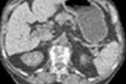
 An 81-year-old woman was scanned for TAVI planning using pitch 1.7 in a single-source CT system, following administration of 60 mL contrast medium at 4 mL/s. Dose length product for entire aorta 147. Images courtesy of Dr. Bernard Bischoff.
Traditionally, TAVI planning with CT has required two separate scans, one focusing on the heart and a second scan of the thorax in order to image the access vessel for the TAVI procedure. But a dual-source CT technique was recently developed to capture all of the relevant anatomy in a single scan.
"In our institution we wanted to see if this dedicated protocol was reserved for dual-source CT, or if it could also be used with single-source, as this would allow many other institutions to utilize this combined protocol," he said.
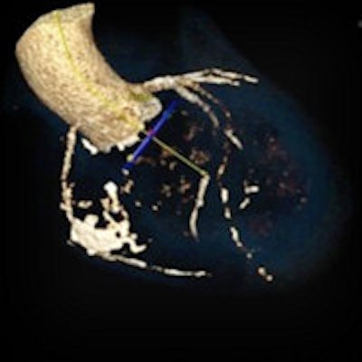
 Transaortic valve implantation planning with dual-source CT as seen on graphic user interface. Images courtesy of Dr. Hans-Christoph Becker.
Aortic valve stenosis is the most frequent valvular disease in Europe, occurring in 2% to 3% of the population age 75 years and older, said Becker, who is a professor of radiology and section chief of body CT at Ludwig Maximilian University.
"It is predominantly a progressive disease, and when symptoms occur they are associated with high mortality," he noted. "Often combined with significant comorbidities, the problem is that a third of patients are not eligible for open heart surgery."
That's where CT for TAVI and transthoracic aortic valve replacement (TAVR) comes in. Less invasive than open heart surgery and better than alternatives, the less invasive technique has been validated in the literature, most recently in a study of 251 patients at 21 surgical centers who were randomized to it or balloon angioplasty, which was found to be inferior.
Five percent of patients undergoing balloon angioplasty suffered a stroke, versus 1.1% undergoing TAVI/TAVR; major vascular complications were seen in 16% of patients undergoing traditional care versus about 1% with TAVI/TAVR, he said.
The comprehensive set of CT measurements required for TAVI planning with CT include the following:
- Assessment of aortic root
- Leaflet calcifications and vegetations
- Annulus sizing and matching specific device
- Size of aortic sinus
- Location of coronary artery ostia
Clinicians must also determine the best imaging plane while minimizing contrast use, determine the best access route for the procedure, and assess comorbidities while ensuring sufficient vessel size, Becker said.
Clinicians have a couple of choices for replacement valves, which differ in the anchor points used. Each comes in three available sizes, including the Corevalve (26 mm to 31 mm) by Medtronic or the Sapient XT (23 mm to 29 mm) by Edwards. A study of 351 patients by Schwartz et al (in press) using one device or the other found that CT predicted vessel size correctly in 84% of all patients.
The study Becker discussed in his presentation, led by his colleague Dr. Bernard Bischoff and published in June, looked at 40 patients with severe symptomatic aortic valve stenosis who underwent CT for TAVI procedure planning. The first 20 consecutive patients (mean age 78 ± 7 years, mean body mass index [BMI] 25.6) were scanned on a second-generation dual-source CT (DSCT) system (Somatom Definition Flash, Siemens Healthcare) using a high-pitch scan mode (pitch value 3.4) and 60 mL of contrast agent.
The second group of 20 patients (mean age 80 ± 7 years, mean BMI 25.1) were examined using a 128-slice single-source CT system (Somatom Definition Edge, Siemens Healthcare), a high-pitch scan mode (pitch value of 1.7), and 60 ml of contrast agent (Imeron 350, Bracco Diagnostics) (International Journal of Cardiovascular Imaging, June 2013, Vol. 29:5, pp. 1159-1165).
The results showed a minor but significant difference in the overall image quality score with lower image quality in single-source CT (3.5 ± 0.6) compared with DSCT (3.85 ± 0.4; p = 0.037). Mean image quality was higher for DSCT (mean score 4.0) compared with single-source CT (mean score 3.5, p = 0.001) for comparing the coronary ostia.
"The image quality was less for the coronary ostia and the ascending aorta, mostly due to the fact we had no ECG triggering for single-source CT, but overall, what can be said was that the images were reliable enough to perform the procedure with single-source CT as well as with DSCT," Becker said.
There was no significant difference in evaluating the aortic valve and annulus, or the image quality of the iliofemoral arteries, and signal intensity and noise did not differ significantly between groups, Becker noted.
| Single-source versus high-pitch dual-source CT angiography (SSCT vs. DSCT) of the aorta for TAVI planning | |||
| SSCT | DSCT | P value | |
| Signal intensity ascending aorta (HU) | 392 ± 102 | 426 ± 116 | 0.425 |
| Image noise ascending aorta (HU) | 18 ± 3 | 18 ±3 | 0.733 |
| Signal intensity iliofemoral (HU) | 338 ± 95 | 356 ± 110 | 0.655 |
| Image noise ileofemoral (HU) | 23 ± 12 | 26 ± 116 | 0.481 |
"We derive the vessel diameters from the dataset and determine the angulation so we can determine the implantation later on," he said. "The workstation also helps us very much to display the access route and the course of the aortic arteries and [determine] whether the catheter and sheath can be pushed through the iliac arteries to perform the procedure at all."
The image quality of high-pitch, whole-body CT angiography with single-source CT was not significantly different from dual-source CT, Becker said, and no significant differences were seen in evaluability, image noise, or signal intensity. Radiation exposure was slightly lower for single-source CT (3.3 ± 0.7 mSv) versus DSCT (4.3 ± 0.8 mSv), and single-source CT also had the advantage of more homogeneous enhancement of vasculature.
"The results are quite remarkable, and I would say there is no significant difference to assess the aortic valve with the single-source CT scanner [or] with dual-source CT," Becker said.