I was talking with a colleague recently about the nature of genius. Apparently on the television in the U.K. at present there is a program about child geniuses, one of whom has a phenomenal memory. Now whilst I would not wish to belittle this achievement, I see genius more as the ability to see reality in a different way from the rest of us, and to do things in a different way. Genius is not really about having a good memory.
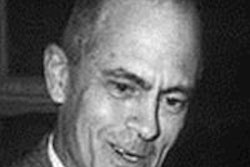
 Gustav Bucky (1880-1963).
Two anniversaries from 1913 are celebrated this year of inventors who saw things differently.1 We have already discussed William Coolidge,2 and he saw a different way of designing an x-ray tube. Gustav Bucky invented a different way of making the x-ray image, and together the two helped define and perfect classical radiography.
It is interesting to observe that when an idea or an invention has reached its time, several people often are working on it independently in the same area. This was true for evolution, with Alfred Russell Wallace and Charles Darwin both working on evolution independently, and for CT scanning, with Godfrey Hounsfield and Allan Cormack working with no knowledge of each other. There are many other examples. The same occurred with the development of the grid.
Experts realized early on that scattered radiation resulted in blurring of the radiographic image. In 1903, Otto Pasche from Switzerland had suggested trying to block the secondary rays by using a slit-like device between the patient and the x-ray tube. However, it was not until 1913 that Gustav Bucky (1880-1963), who was a German radiologist, designed a honeycombed and grid-like diaphragm, which he placed between the patient and the x-ray plate.3
This metal lattice had individual cells that allowed the primary x-rays from the focal spot of the x-ray tube to pass directly through to the film. Secondary scattered radiation was blocked by being absorbed by the metal strips. The major disadvantage was that the grid was visualized on the x-ray plate and obscured some of the findings.
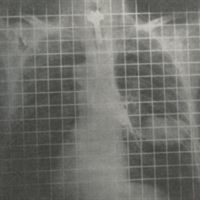
 A Bucky grid made in 1913 in Germany by the Siemens & Halske Company.
 An early bronchogram using the stationary crisscross Bucky grid with grid lines visible.
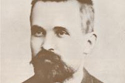
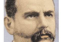
Bucky came up with a possible solution, which was to move his grid so the shadows of the strips were no longer visible. It is interesting that quite independently in 1917 Eugene Caldwell (1870-1918) from New York had the same idea of moving the grid to remove the shadows. A third investigator, Hollis Potter (1880-1964) from Chicago, was unaware of the previous publications and presented his ideas at meetings of the American Roentgen Ray Society (ARRS) in 1915. Potter described a diaphragm that he adapted for fluoroscopy and consisted of a rotating circular disc with radiating strips to absorb the scatter.
 Interception of scattered rays by a grid.
These early grids with the crisscross pattern were neither successful nor popular. However, Potter was aware that a moving wire did not cast a shadow on an x-ray plate, and so he realized this would also happen if a lead strip that moved uniformly across the beam was used. Potter created a series of parallel strips, and although this should only have absorbed a portion of the scattered radiation, Potter found not only was the total amount absorbed similar to that of the crisscross Bucky grid, but also it was much easier to manufacture.
 The Potter-Bucky diaphragm.
This new device was marketed as the Potter-Bucky diaphragm and was announced in February 1917 at the ARRS.4 Potter demonstrated the use of this apparatus to show the lumbar spine, hips and pelvis, and renal calculi. It is famously recorded that at one meeting after Potter had shown his work, one radiologist was so surprised by the quality of the images that he accused Potter of touching up the negatives!
There was a certain delay in making the apparatus commercially available because of discussions about the priority of the discovery. However, in 1921 the Potter-Bucky diaphragm was finally marketed and was immediately accepted by the radiological profession. Whilst the image of the pelvis from 1921 is excellent, it should be noted that the exposure time was 12 seconds!
One effect of the new grid was to hasten the demise of x-ray glass plates because with the new Potter-Bucky diaphragm, larger films could be used with limited effects from scatter and the consequent blurring. Scatter was more the problem with larger films than smaller films and traditionally radiologists were restricted to smaller plates. The smaller the plate, and therefore the volume irradiated, the less scatter was generated. The reason that a cone had been introduced over the x-ray tube was to reduce the irradiated volume and therefore the scatter.
It is worth recording that at the end of World War I, the U.S. government confiscated many German patents, including that of Bucky. His patent ran out in 1933, and whilst Bucky should have received an estimated $4 million in royalties, he never received anything. Bucky himself emigrated to the U.S. in 1923, bur he was always concerned that Potter was so much associated with the invention. While Bucky made the initial invention, it was Potter's moving grid with longitudinal lead strips that was practically and commercially popular.
Since the 1920s, the idea of the grid has continued to evolve, including development of focused grids and very fine grids. Therefore, it was the combined developments in 1913 of the Coolidge tube and the Bucky grid that contributed hugely toward the flourishing of classical radiography. As such, we must salute both of their geniuses. And we should also look at what we are doing today and ask, "Can we do things differently and better?"
 Normal pelvis from 1921 using the Potter-Bucky diaphragm and a Coolidge tube.
Dr. Adrian Thomas is chairman of the International Society for the History of Radiology and honorary librarian at the British Institute of Radiology.
References
- Thomas AMK, Banerjee AK. The History of Radiology. Oxford: Oxford University Press; 2013.
- Thomas AMK. "Reflections on the incredible life of William David Coolidge." AuntMinnieEurope.com. http://www.auntminnieeurope.com/index.aspx?sec=sup&sub=xra&pag=dis&ItemID=607705. 8 February 2013.
- Moehring HG, Grids. In: Bruwer AJ, ed. Classic Descriptions in Diagnostic Roentgenology. Springfield, IL: Charles C Thomas; 1964.
- Holbeach CH. The Potter-Bucky Diaphragm. The Journal of the Röntgen Society. 1921;17:179-181.
The comments and observations expressed herein do not necessarily reflect the opinions of AuntMinnieEurope.com, nor should they be construed as an endorsement or admonishment of any particular vendor, analyst, industry consultant, or consulting group.