Imaging the femaile pelvis is possible using contrast-enhanced 7-tesla T1-weighted MR imaging, according to German researchers, who also demonstrated the image impairment associated with T2-weighted MRI in their study published in European Radiology. Their preliminary results show that 7-tesla MRI of the pelvis is a promising but challenging imaging technique.
MRI of a woman's pelvis has been described as the most accurate, noninvasive imaging investigation for determining cervical carcinoma size and uterine extension. The modality is also good for evaluating recurrent disease. Imaging at 1.5 tesla is currently considered the standard for pelvic MRI; however, it has limitations in its diagnostic accuracy for assessing tumor extent and/or parametrial invasion, mainly because of its low signal-to-noise ratio (SNR), according to the researchers.
An increase in SNR, and consecutively the contrast-to-noise ratio, can be achieved by an increase in the magnetic field strength. Numerous studies have demonstrated the diagnostic potential of 3-tesla pelvic MRI by transforming the associated gain in SNR into higher spatiotemporal resolution.
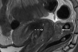
 T2 turbo spin-echo imaging (A) of the female pelvis revealing good quality delineation of the zonal anatomy of the uterus. 2D FLASH post-contrast imaging (B) shows lack of enhancement of the ovarian cysts (arrows). The thin arrow in figure B marks a residual inhomogeneity artifact in the right gluteal muscle. All images courtesy of Dr. Lale Umutlu.
Ultrahigh-field (7-tesla) MR systems have been successfully implemented for in vivo human imaging and are becoming increasingly established for structural and functional brain imaging at research sites worldwide, wrote Dr. Lale Umutlu, from the department of diagnostic and interventional radiology and neuroradiology at University Hospital Essen, and colleagues.
"Recent technical advances have been introduced to counter some of the particular challenges of imaging at 7[-tesla], and scientific research has arisen to broaden possible areas of application from neuroradiology to body imaging," they added (Eur Radiol, 4 May 2013). "Starting out with ultrahigh-field whole-body landscape imaging, the focus of studies has shifted toward the implementation of dedicated abdominal MRI, as well as morphological and functional cardiac MRI."
With ultrahigh-field MRI being increasingly used for body applications, Umutlu and colleagues sought to investigate the feasibility of contrast-enhanced ultrahigh-field MR imaging of the female pelvis.
Ten healthy women volunteers were examined on a 7-tesla whole-body MR system (Magnetom 7-tesla, Siemens Healthcare) utilizing a custom-built eight-channel transmit/receive radiofrequency body coil. The protocol included T1-weighted fat-saturated 2D spoiled gradient-echo (FLASH), dynamic T1-weighted fat-saturated 3D FLASH, and T2-weighted turbo spin-echo (TSE) sequences.
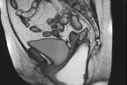
 Nonenhanced (A) versus post-contrast (B) 2D FLASH imaging of the pelvis. Notice the equivalently high-quality delineation of the arterial and venous vasculature (arrows).
For qualitative image analysis of pelvic anatomy, the researchers assessed uterine zonal anatomy and image impairment due to artifacts using a five-point scale. For quantitative analysis, contrast ratios between the junctional zone and myometrium were obtained for T2-weighted MRI.
The researchers found 2D FLASH MRI offered the best overall image quality (mean contrast-enhanced 4.9) and highest tissue contrast (mean contrast-enhanced 4.7), while T2-weighted TSE imaging provided moderate to high conspicuity of the uterine zonal anatomy, with mean scores ranging from 3.5 for endometrium to 4.65 for myometrium. Overall image impairment was rated strongest for T2-weighted MRI (2.9) and least for 2D FLASH MRI (mean 4.2).
"These preliminary results demonstrate that contrast-enhanced 7-[tesla] MRI of the pelvis is a promising, yet also challenging imaging technique, showing considerable differences in the current technical viability of T1- and T2-weighted imaging," Umutlu and colleagues wrote.
T1-weighted fat-saturated 2D FLASH MRI resulted in good image quality, providing excellent tissue contrast and high spatial resolution and revealed the potential for ultrahigh-field perfusion MRI, enabling further evaluation of possible pelvic pathologies.
T2-weighted TSE MRI showed high-quality delineation of specific uterine anatomy with comparable contrast to lower field strengths, yet remained challenging because of limitations associated with ultrahigh magnetic field strength, they wrote.
One such limitation is an obstacle posed by specific absorption rate (SAR) restrictions, as the energy deposition in tissue increases with the square of the magnetic field and of the flip angle, as well as being proportional to the duty cycle of the radiofrequency (RF) pulse.
"This is especially problematic for the adequate acquisition of RF intense turbo spin-echo and fat-saturated sequences," the researchers wrote. "To mitigate the associated SAR increase, parallel imaging and a modified T2-weighted TSE sequence with variable refocusing flip angles were applied in this study."
However, the diagnostic value of this sequence type was strongly limited, as most images fell short of diagnostic quality, they noted.
Increasing RF inhomogeneity with increasing field strength also posed challenging, as inadequate homogeneity of the RF magnetic field impairs the generation of accurate refocusing pulses, which are a prerequisite for high-quality TSE imaging.
"To mitigate the effects of these variations in magnetic field uniformity, dedicated B1 shimming was performed with a custom eight-channel RF shimming setup to reduce B1 artifacts in the region of interest," the researchers wrote. "Nevertheless, minor impairment due to variations in B1 uniformity remained as demonstrated in mild differences in anterior versus posterior fat signal intensities; these might be addressed with an RF shimming system with a higher degree of freedom in future studies."
Recent updates on 7-tesla MRI of the pelvis include further work and improvement of fat saturation in the dynamic T1-weighted 3D sequences, as well as coping with the limitations in T2-weighted MRI, Umutlu said in an interview with AuntMinnieEurope.com.
"We have just set up a protocol for the first patient studies for evaluation of the diagnostic competence of 7[-tesla] pelvis imaging in comparison to 3[-tesla] MRI for assessment of female pelvic tumors (e.g., cervical cancer) and will start them in the near future," she said.
Future studies may also have larger cohorts including patients with gynecologic disease as well as comparison trials to lower field strengths.