Anterior cruciate ligament (ACL) tears are not isolated events, but only the most obvious sign of a complex knee injury involving multiple structures. Awareness of the broad spectrum of MRI appearances at common sites of associated injury is vital, according to a multinational group of musculoskeletal experts.
"In day-to-day practice, the distinction between normal and pathologic appearance of each structure at the knee can be difficult to make, particularly in adolescents," noted lead author Dr. Jacob Jaremko, from the department of radiology and diagnostic imaging at the University of Alberta, Edmonton, Canada. "A child with an ACL tear has a lifetime of loading ahead, and for optimal planning of surgical treatment and prediction of long-term clinical outcome, the radiologist should report the ACL tear with an integrated description of associated findings implying the likely injury mechanism."
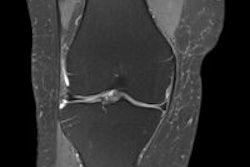
 Distal rupture of the medial collateral ligament (arrow) on coronal short-tau inversion recovery of a 15-year-old boy. All images courtesy of Drs. Jacob Jaremko and Zachary Guenther.
Along with colleagues from Ghent University Hospital in Belgium and Mater Misericordiae University Hospital in Dublin, he has shared his experiences of imaging of ACL tears in an article in Insights into Imaging, posted online on 9 May. The authors are concerned that insufficient attention is given to children and adolescent injuries.
In these cases, radiologists must be aware of tibial spine avulsion instead of ACL rupture, a thin but intact ACL, poor visualization of posterolateral corner structures, and differentiation between incompletely closed physis and impaction fracture. The posterior lateral meniscus is particularly complex and should be studied in three regions (posterolateral corner, meniscofemoral ligament attachment, and bony attachment), and if an ACL tear is detected, the associated findings described here should be specifically sought and, conversely, observing one of these findings should increase the suspicion for an ACL tear, they stated.
Among the key points to bear in mind are:
- The ACL in children normally appears thin or attenuated, while thickening and edema suggest a tear.
- Displaced medial meniscal tears are significantly more common later postinjury than immediately.
- The meniscofemoral ligaments merge with the posterior lateral meniscus, complicating tear assessment.
- Tibial plateau impaction fractures can be difficult to distinguish from a partially closed physis.
- Axial MR sequences are more sensitive/specific than coronal for diagnosis of medial collateral ligament injury.
ACL injury usually occurs with axial rotation in the valgus near full extension, the authors explained. In cases of ACL rupture, MRI findings can be obvious and important to patient management, subtle but clinically important (lateral meniscus posterior attachment avulsion), obvious and unimportant to management (femoral condyle impaction injury), or subtle and possibly important (medial meniscocapsular junction tear).

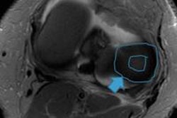
 Left: Bucket-handle tear of the medial meniscus in a 16-year-old girl. Coronal proton-density image demonstrates irregular, torn, medial meniscal body that is smaller than normal (white arrow), and the flipped portion of the medial meniscus that appears as an unexpected low signal structure below the posterior cruciate ligament in the intercondylar region. Right: Femoral condyle impaction fracture in a 15-year-old girl. Sagittal proton-density image shows cortical depression (arrow) with surrounding edema. Note the associated complex tear of the posterior horn of the lateral meniscus.
In younger patients, the normal ACL can appear quite attenuated, and a recently torn ACL is usually visibly thickened and edematous, and a thin and appropriately oriented ligament without edema is likely intact, they stated. In the last 110 cases at the University of Alberta, 7/48 (15%) read by musculoskeletal radiologists and 26/62 (42%) read by pediatric radiologists were initially reported as high-grade partial-thickness ACL tears, while at later surgery all but one of these tears (99%) was described as complete.
"Given the time delay between injury and surgery, it is possible that partial tears evolved to complete tears at surgery; however, the distinction between complete and partial tears is well known to be limited in reliability on MRI," Jaremko et al wrote. "One challenge is that the torn ACL may form scar tissue that can mimic a partially intact ligament. A tear that appears to involve most of the ACL fibers is likely to be functionally complete, and we encourage reporting the tear in this fashion."
Awareness of the spectrum of secondary findings and the features distinguishing them from normal variation can aid in accurate assessment of ACL tears and related injuries, enabling effective treatment planning and assessment of prognosis, they concluded.