Into the torrent of research from Europe that applies imaging technology to forensic tasks comes a new Austrian study that uses a computer algorithm to pinpoint both the age and sex of skulls.
The computerized method outperformed both visual assessment and a conventional technique called the Knussmann scheme that does the same task based on bone surface curvature. In fact, the algorithm reached an accuracy of 100% in four bones in the skull, and 75% to 100% in two other bones, compared with a broad range of 4.2% to 100% accuracy for the Knussmann scheme.
"The results of this promising method give us the capability of using bone shape for age estimation and an analysis of surfaces changes during life," said Dr. Alexander Vlcek from the Medical University of Vienna. However, he stressed new techniques are needed for forensic determination of the sex and age of human bones because existing techniques are both inaccurate and time-consuming.
Visual assessment of bone shape has not been fully validated for the estimation of sex and age, he explained. Osteometry and standard measurements have yielded accuracies of about 83% to 100%. The Knussmann scheme is the standard method used for decades to investigate the sex based on the surface expression of bone, but it is subjective and imprecise. It has a smoothing effect, so convex surfaces may be overlooked, noted Vlcek.
Could a surface analysis program commonly used in virtual colonography computer-aided detection (CAD) schemes be applied to the job of determining the sex and age of human cranial bones? Vlcek, along with Dr. Fabian Kanz, Wolfgang Weninger, PhD, Dr. Daniele Risser, and others, aimed to find out, and they presented their findings at ECR 2013. They looked at the feasibility of using a computerized method incorporating shape index (SI) and surface analysis to estimate the sex of skull remains based on age-associated changes to the skeleton on macerated skulls, and then they compared them to subjective assessments applying the osteoscopic Knussmann scheme.
"In the prestudy, five individuals (three males and two females) were taken just to see if there was any chance with the shape index," Vlcek said. "In the main study, the same procedure was repeated on 24 skulls and the surface analysis was added."
The prestudy examined whether Koenderink's shape index -- a technique commonly applied in virtual colonoscopy CAD schemes -- could be applied to the cranial bones of the five specimens. The main study examined the skulls of 24 Europeans, including 12 men (aged 23 to 77 years, mean 45 years) and 12 women (aged 22 to 75 years, mean 46 years).
All analyzed macerated skulls were taken from historical collections, and the researchers attempted to keep the size as high as possible. The causes of death ranged from malnutrition to crime; for inclusion in the study the sex and age had to be known and the full specimen had to be available, he said.
The skulls were scanned using a 64-detector-row CT unit (pitch 0.64) and modeled using AMIRA 5.4 software to calculate the percentage deviation of local concavity or convexity in five sex-specific traits in the frontal bone, zygomatic bone, mastoid process, planum nuchale, and mentum as measured with the shape index, Vlcek said.
Based on the results of this analysis, the group defined different surface shapes and two indices, and sex-differentiating formulas were developed using regression analysis. For determining the surface shape, the range between convex and concave was divided into nine surface shapes, and regression analysis was used to implement the nine surface shapes as percentage values. Results in the 24 skulls were compared between the computer algorithm and visual assessment.
The prestudy showed Koendrink's shape index can be applied with high accuracy to skulls, and the reported accuracy was 67.7% to 100% in five bones, with better results in women's skulls, he noted.
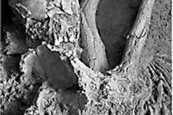
 Prestudy shape index. All images courtesy of Dr. Alexander Vlcek.
Results in the 24 skulls showed that computer-assisted shape index calculations are superior to visual assessment across all seven bone types examined, with accuracies of 66.7% to 100% versus 0% to 91.7% for visual assessment, Vlcek said.
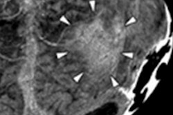
 Shape index is superior to visual assessment.
The study also demonstrated changes in bone shape from childhood to adulthood can be plotted -- mainly on the convex surfaces of the bone -- in the frontal bone and mastoid process, he said.
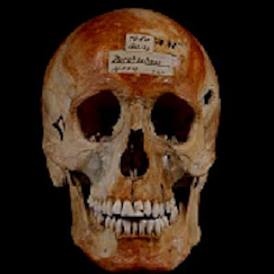
 Surface changes between childhood and adulthood can be plotted, with larger changes seen in women.
The limitations of the results lie in the small sample size, the fact that age was the only trend analyzed, and the rather crude segmentation process, which will be refined in future studies that will also include more samples, according to Vlcek.
Shape index is independent from light options and smoothing effect, and the computerized method is promising for sex estimation and analysis of surface changes, he said.