Free breathing by patients can reliably reduce uncertainty in tumor position caused by respiratory motion during MRI-guided radiation therapy procedures on the kidney, according to Dutch researchers.
A team from the University Medical Center Utrecht in the Netherlands found that free breathing decreased kidney position variability more than breath-hold techniques. They report their findings in an article published in the 7 April issue of Physics in Medicine and Biology.
The capabilities of hybrid MRI radiotherapy modalities may make it possible to delivery radiation dose to treat cancers for which surgery is usually the first line of treatment. But when a cancer is in an organ subject to motion, such as a kidney, this can be challenging, according to the group led by lead author Dr. Mette Stam of the department of radiotherapy.
The current treatments for individuals diagnosed with kidney cancer, whether a radical or partial nephrectomy, cryoablation, or radiofrequency ablation, are associated with high complication rates due to their invasive nature. Radiotherapy is currently used when surgery is not an option. The precision offered by image-guided radiation therapy (IGRT) has the potential to target renal cell carcinoma, but uncertainties in tumor position caused by respiratory motion need to be overcome.
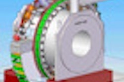
As a result, a team at the university, where a MRI linear accelerator is in development, sought to evaluate the techniques of free breathing and nonguided breath-holds to determine their efficacy (Phy Med Biol, 7 April 2013, Vol. 58:7, pp. 2235-2245). The new IGRT prototype being developed by Elekta and Philips Healthcare at the university is a combination of a 6-megavolt linac and a 1.5-tesla MRI scanner.
The capability of providing MRI during the irradiation process will allow radiation therapists to monitor the position of a tumor with real-time imaging and adapt the treatment to the patient, according to the authors. To determine the stability and reproducibility of the kidney position, the research team recruited 15 patients with renal lesions to be imaged while using two breathing techniques.
Free breathing
Both sagittal and coronal dynamic scans of the kidney were acquired during free breathing. Stam and colleagues determined the mean and standard deviation of the expiration peaks after they were determined. They also created a linear fit of the kidney positions during expiration, and these calculations helped the research team determine the reproducibility of the kidney position.
A gating window was also calculated to determine the percentage of time in which the kidney was inside it. The authors positioned the gating window symmetrically around the average expiration position of the kidney in the first five breathing cycles. They made calculations for gating windows that varied in size between one and 20 mm.
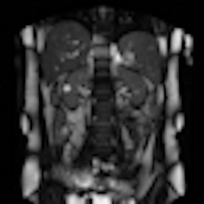
 Left: Coronal and Right: sagittal time frame in 2D cine MRI. Image reproduced with permission from IOP Publishing.
The researchers concluded that the cranial caudal direction was the largest component of kidney motion. The smallest component proved to be the right-left direction, where a variation smaller than 1 mm over all breathing cycles was identified in one-third of the patients.
Expiration positions during free breathing were reproducible in 80% of the patients within 2 mm, and for more than half of the patients the reproducibility was smaller than 1 mm. Efficiency increased as the gating window increased, according to the authors.
Breath-hold technique
The 15 patients performed ten consecutive breath-holds, five for coronal scans and five for sagittal scans. The researchers positioned an acceptance window on the reference kidney positioned, and determined the percentage of time that the kidney was within it. They also investigated whether correcting the position of the acceptance window for baseline shifts between breath-holds would improve the treatment efficiency. For both, the size of the window varied between 1 mm and 20 mm.
The researchers determined that treatment efficiency is improved when the acceptance window is repositioned at the start of each breath-hold. They did not estimate the time needed to prepare a patient for a breath-hold, but did record the time between breath-holds to be an average of 43 seconds.
The variability of the expiration breath-hold position provided to be bigger than the variability of the expiration position during free breathing for all of the patients. Treatment efficiency also was higher during free breathing than with the breath-hold technique. Additionally, patients were more comfortable with the free breathing technique.
The researchers plan further investigation using audiovisual feedback to evaluate gating efficiency and reproducibility of the breathing pattern during free breathing.