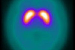
Researchers from Japan have collected a prestigious award for what is thought to be the first-ever study directly comparing postmortem neuromelanin MR imaging (NmMRI) and neuropathological findings in elderly patients.
"Our results show that signal intensity in the SNc (substantia nigra pars compacta) is closely related to the quantity of neuromelanin-containing neurons as shown by the direct correlation between neuromelanin imaging and neuropathological findings," noted Dr. Shinichiro Kitao, from the division of radiology in the department of pathophysiological and therapeutic science at Tottori University, Yonago, Japan, who received one of only five Magna Cum Laude awards given for e-posters presented at ECR 2013.
Neuromelanin is a byproduct of the synthesis of monoamine neurotransmitters such as noradrenalin and dopamine, and is mainly expressed in neurons of the locus coeruleus (LC) and SNc. Neuromelanin can induce paramagnetic T1-shortening effects when combined with metals such as iron and copper, but T1-weighted imaging has failed to depict neuromelanin-generated contrast, presumably because of the negligible T1 difference between neuromelanin and brain tissue, according to the authors.
Parkinson's disease is characterized by severe loss of dopaminergic neurons and neuromelanin, and pathologically, dementia with Lewy bodies (DLB) closely resembles Parkinson's disease. Patients with DLB are characterized by the diffuse presence of Lewy bodies, cytoplasmic inclusions composed principally of alpha-synuclein, in both subcortical and cortical areas of the brain, whereas Parkinson's disease patients have Lewy bodies in the subcortical areas of the brain, mainly the SNc and LC, they explained.
On neuromelanin-contrast imaging, paramagnetic T1-shortening effects of neuromelanin-containing neurons are thought to be closely related to the high signal intensity of the SNc. In addition, the iron content of the SNc is very high (approximately 20 mg/100 mg tissue) and it increases with age. Therefore, the possibility that a high iron concentration in the SNc may cause T1-shortening effects has been suggested in neuromelanin-contrast imaging.
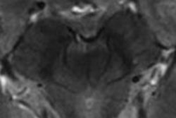
Kitao and colleagues retrospectively analyzed MR brain images in three autopsy-proven cases: an 81-year-old man with normal neuropathological findings, an 81-year-old man with DLB, and a 68-year-old woman with Parkinson's disease. NmMRI of 10% -formalin-fixed autopsied brains was obtained using a 3-tesla imaging system (Signa Excite HD, GE Healthcare). For MR-pathologic direct correlation, neuropathological examinations were performed in all cases.
Postmortem NmMRI of the midbrain showed diffuse hyperintense areas in the SNc, and the signal intensity of the SNc was higher than that of the SC. Myelin staining of the SNc was the same as that of the SC, whereas strong myelin staining was seen in the red nucleus, medial lemniscus, and cerebral peduncle. Diffuse ferritin deposition in the SNc was fainter than that in the red nucleus, medial lemniscus, and cerebral peduncle. Histologically, the diffuse hyperintense area in the SNc reflected well-preserved neuromelanin-containing neurons, whereas the hyperintense area with multiple hypointense spots in the SNc reflected neuromelanin-containing neurons with bundles of myelinated nerve fibers and dilated perivascular spaces.
In the normal control case, hyperintensity with multiple hypointense spots was seen in the SNc, the authors stated. Histologically, these hypointense areas reflected dilated perivascular spaces and bundles of myelinated fibers in the SNc. These structures were also seen in cases with DLB and Parkinson's disease. Decreased signal intensity reflecting bundles of myelinated fibers seems to be influenced by formalin fixation in postmortem T1-weighted images, a finding that is not seen in antemortem T1-weighted images.
"We evaluated whether iron deposition contributes to high signal intensity in NmMRI. In all cases, NmMRI of the midbrain showed hypointensity in the red nucleus and white matter such as the medial lemniscus and cerebral peduncle. Furthermore, diffuse ferritin deposition in these regions was stronger than in the SNc. Therefore, increased signal intensity in the SNc seems to not be influenced by iron deposition," they added.
The authors admitted the study has several limitations. First, they did not evaluate signal changes with iron imaging such as spin echo T2-weighted imaging, T2*-weighted imaging, and susceptibility-weighted imaging. Instead, iron deposition in tissue was directly evaluated using ferritin histochemistry. Iron deposition apparently did not contribute to the high signal intensity on NmMRI. Second, this study involved a small number of subjects. Third, the LC was not evaluated in this study, although this structure is usually involved in Parkinson's and DLB.
Inspite of these limitations, signal intensity in the substantia nigra is closely related to the quantity of neuromelanin-containing neurons and is apparently not influenced by iron deposition, based on the direct correlation between NmMRI and neuropathological findings, the team concluded.