Errors in radiology are common, particularly when it comes to the review of digital x-ray and CT images. But most discrepancies occur in a small number of areas, and focused review of these areas can significantly reduce error rates, according to prize-winning research from a U.K. teaching hospital.
"Within radiology, our errors are saved in indelible electronic ink for the entire world to see," noted Dr. Jonathan Weir-McCall, a radiologist at Ninewells Hospital, University of Dundee, U.K. "Medicine has long functioned as a large number of autonomous individuals with a blame culture that lags behind the reasoned safety culture present in other walks of life such as the aviation industry. However, this ease of identifying errors should be seen as a tool to create opportunities to improve, rather than as a cross upon which to crucify."
Radiological societies in the U.S., U.K., Australia, and elsewhere recommend discrepancy meetings in which to discuss errors, and there is a growing acceptance that review of cases in an organized meeting with constructive feedback can reduce error rates, he explained in an RSNA 2012 e-poster that received a certificate of merit.
Ninewells is a 995-bed teaching hospital covering a population of around 450,000, and it has operated a monthly radiological error and discrepancy review meeting since 2007. Cases of clinical importance/educational value are discussed to guide and improve practice. Mammography and interventional procedures have their own discrepancy process, so were excluded from the study. Discrepancies were split into system of origin, before then being subclassified and split according to type of error.
To date, the radiology department has built up a database of 561 errors, 116 of which originated from cases in the central nervous system (CNS), 99 in the thorax, 290 in the abdomen, and 119 in the musculoskeletal (MSK) system; some errors originated from more than one system, hence the apparent statistical anomaly.
The type of errors were: false positive (28), false negative (381), misclassification (104), technical (13), and communicative (35). A total of 103 errors were related to the use of plain film, 14 to fluoroscopy, 43 to ultrasound, 320 to CT, and 81 to MRI, according to Weir-McCall.
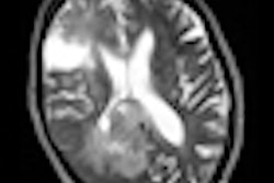
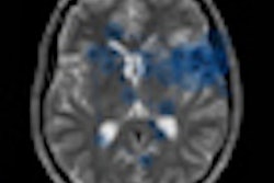
The five most common CNS errors (vascular, infarct, cerebral mass, bone, cerebrospinal fluid) accounted for 57% of CNS reporting discrepancies. In thoracic cases, the five most common errors (nodule, bone, vascular, mediastinal, lymph nodes) account for 69% of thoracic reporting discrepancies.
"Despite knowledge of the chest x-ray review areas, the majority of the missed nodules were in the traditional review zones. This is not surprising given the many overlying structures in these zones, making them difficult to analyze," he wrote.
All but one of the 14 vascular discrepancies were due to missed pulmonary emboli. Of the 13 missed pulmonary emboli, only one occurred on a dedicated CT pulmonary angiogram.
The five most common abdominal errors (vascular, colonic, renal, liver, pancreas) accounted for 54% of thoracic reporting discrepancies. On CT, the kidneys are best visualized on coronal images because lesions can often be missed on axial slices, advised Weir-McCall and his co-authors. The liver, on the other hand, is a large organ within which small subtle lesions can hide, so multiplanar reformatting can help visualize lesions that are difficult to see on only one plane. In the pancreas, satisfaction of search and failure to review previous images are common pitfalls in accurate image interpretation, they pointed out.
In MSK cases, peripheral limb fractures are often subtle and easy to miss, especially in polytrauma patients with distracting injuries that require more immediate attention. Also, benign bone lesions are often automatically assumed to be malignant on staging CT examinations. Finally, lesions to the periphery of a CT/MRI scan are easily missed, and clinically unsuspected MSK infection is easy to miss, especially when it affects a peripheral site on an MRI or CT scan, the authors concluded.