A new reconstruction algorithm increases the accuracy and diagnostic performance of dual-source CT angiography (CTA) by raising the temporal resolution of images acquired at slow gantry rotation speeds, making it suitable for application in less costly scanners and for special cases such as obese patients, a German-U.S. research project has found.
The Temporal Resolution Improvement Method (TRIM, Siemens Healthcare) algorithm uses an iterative technique to reconstruct images from projections less than 180°, and employs a histogram constraint to prevent limited-angle artifacts, noted authors from the University of Heidelberg in Germany and the Medical University of South Carolina (MUSC) in the U.S., along with its developer, Siemens Healthcare.
Use of the TRIM algorithm significantly improved temporal resolution in a cohort of 11 obese patients (7 men; mean 67.2 ± 9.8 years) with a mean body mass index (BMI) of 36 kg/m2. Average heart rate during imaging was 60 ± 9 beats per minute (bpm).
 |
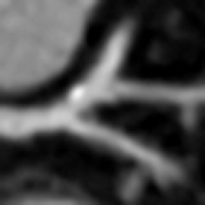
| Compared with filtered back projection reconstruction (above), use of the TRIM algorithm improves temporal resolution and renders a more diagnostic image. Images courtesy of Dr. U Joseph Schoepf, MUSC. |
 |
"Despite cardiac CT image acquisition with slower gantry rotation, the temporal resolution improved reconstruction algorithm evaluated here provided more diagnostically adequate image quality for coronary artery assessment than the standard FBP [filtered back projection] reconstruction algorithm," wrote Dr. Paul Apfaltrer from the University of Heidelberg and MUSC, and colleagues in an article-in-press in the European Journal of Radiology (November 5, 2012).
Artifacts from cardiac motion can limit the accuracy of coronary CTA (CCTA), and this problem was the impetus behind the 2006 introduction of dual-source CT, which increases temporal resolution of 75-83 msec and as a result the robustness of CCTA for ruling out coronary artery disease. Changing the reconstruction algorithm to extract higher resolution images is another way to improve temporal resolution, and was the aim of this study.
TRIM algorithm
The TRIM algorithm works as follows: A short scan FBP reconstruction of the image data is created, resulting in a temporal resolution equal to half the rotation time. Then the 180° images are partitioned into several smaller regions, and its histogram is computed for each region. Next the iterative simultaneous algebraic reconstruction technique (SART) at the heart of the algorithm is applied using only a subset of the short-scan projection data, the authors explained.
"Before every SART iteration, an additional step is undertaken where the HU values of each pixel are adjusted according to the histogram of the low temporal resolution image: Pixels with a Hounsfield unit (HU) value close to a maximum of the histogram are left unchanged; pixels with a HU value far from any maximum are adjusted slightly toward the closest maximum," they wrote. Finally, using a motion detection technique the TRIM image is combined with the 180° FBP image.
In the study, all patients underwent dual-source CTA (Definition Flash, Siemens Healthcare) using the XXL protocol for obese patients and 0.6-mm collimation, 120 kV, 175-426 mAs, and 500 msec gantry rotation speed. Before scanning, patients received iodinated contrast (triphasic protocol, 70 mL of iopromide, 370 mg/mL of iodine), sublingual nitroglycerin, and beta-blockers to reduce the heart rate.
The image data were transferred to a 3D workstation (Circulation, Syngo MultiModality Workplace, Siemens Healthcare) and reconstructed using a temporal resolution of 250 msec using both FBP and resolution of 200 msec using the TRIM algorithm. Two experienced readers examined the images independently, measuring contrast attenuation and contrast-to-noise-ratio (CNR) in the ascending aorta, and rated the presence and severity of coronary motion artifacts on a four-point Likert scale. Mean effective dose for the procedure was 13.5 ± 4.6 mSv, and the average heart rate was 60 ± 9 bpm. The results in 110 coronary artery segments were as follows:
There were no significant attenuation differences between the FBP and the TRIM reconstructions in the ascending aorta (392 ± 70.7 versus 397 ± 70.1 HU, p > 0.05), and there were no significant differences in contrast attenuation.
But there were differences in motion artifacts. Coronary CTA studies reconstructed with the TRIM algorithm showed a significantly lower median severity score of coronary motion artifacts compared with the FBP reconstruction (2.0 versus 2.5, p = 0.007), the authors reported.
FBP showed 10% of segments with no artifacts, 42% with minor artifacts, 41% with moderate artifacts, and 7% with severe artifacts, for a nondiagnostic rate of 7%. In contrast, the TRIM algorithm showed 15% of segments with no artifacts, 71% with minor artifacts, and 9% with moderate artifacts. Five of these segments were given a Likert score of 4, and were considered nondiagnostic, the group reported.
Image quality based on coronary motion artifacts with FBP versus or TRIM algorithm reconstruction
|
Despite the slow gantry rotation for the cardiac CT acquisition, the TRIM algorithm offered more diagnostically adequate image quality for evaluating the coronary arteries than standard FBP reconstruction techniques, Apfaltrer and his team wrote. For cardiac CT, 180° (half a gantry rotation) of projection data are traditionally needed to reconstruct diagnostic images; the temporal resolution of single source CT scanners has historically been defined as half the gantry rotation speed.
"The primary approaches for improving temporal resolution at cardiac CT thus far have mostly involved hardware optimization, with ever faster gantry rotation and/or dual-source data acquisitions," Apfaltrer and colleagues wrote. "However, the associated system complexity of high-end scanners is costly and such systems are not ubiquitously available. The majority of all cardiac CT studies are currently still performed on the installed base of 64-slice CT systems, with limited temporal resolution."
Even with the latest, fastest scanners with the highest temporal resolution, certain patient scenarios favor the use of slower gantry rotation speeds, such as in obese patients to boost the photon flux through the patients and offset the photon starvation that would otherwise occur at fast gantry rotation speeds, they wrote.
"The penalty of this approach, of course, is the trade-off between a higher contrast/noise ratio [and] slower temporal resolution," they stated. Various image reconstruction algorithms have been pursued to improve temporal resolution and reduce cardiac motion artifacts. But the TRIM algorithm used for this study may be more robust and easier to implement than previous methods, such as temporal resolution improvement with prior image constrained compressed sensing (TRI-PICCS) and multisegment reconstruction.
"Our results suggest a reduction of temporal resolution to values below the half-rotation limit, which translated into a significant reduction in the severity scores of coronary motion artifacts compared to the traditional FBP reconstruction," the authors noted. "In particular, there were significantly fewer segments showing moderate motion artifacts and more segments with only minor motion artifacts. This motion artifact reduction due to temporal resolution improvements may thus improve the diagnostic value of slower gantry rotation systems in the detection and exclusion of coronary artery stenosis with coronary CTA."
Among the limitations, larger studies are needed to confirm the finding, the group wrote. In addition, evaluation of the advanced image reconstruction algorithm does not show improved diagnostic sensitivity, which also will need to be demonstrated.
"The algorithm evaluated here allows clinically adequate imaging of the coronary arteries by restoring acceptable temporal resolution despite slower gantry rotation speeds, and might therefore improve the performance of slower gantry rotation systems for noninvasive imaging of the coronary arteries with CTA," the group concluded.