Asbestos-related disease remains an important issue due to the long latent period between environmental or occupational exposure and clinical manifestation, and radiologists must be able to distinguish between common, asymptomatic conditions and serious, debilitating or malignant cases, according to U.K. research.
"Pleural plaques are virtually pathognomic of asbestos exposure but noncompensable. Other thoracic manifestations of asbestos exposure are less specific and may be seen in nonexposed individuals," noted Dr. D. Martin and colleagues from the department of radiology at University Hospital Llandough in Cardiff, U.K. "In assessment of asbestos-related claims, detailed occupational or environmental history is essential. Other potential causes of thoracic manifestations should be excluded."
Thoracic manifestations of asbestos exposure include benign pleural disease (plaques, effusions, diffuse pleural thickening), benign parenchymal disease (rounded atelectasis, asbestosis), and malignancy (mesothelioma and lung cancer).
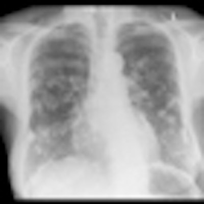
 Digital chest x-ray of female patient shows multiple calcified pleural plaques caused by asbestos exposure. Plaques are also seen along the diaphragm.
Effusions in benign pleural disease are usually seen within 10 years of exposure, and they are often asymptomatic, small (less than 500 mL), and exudative. In many cases, they resolve spontaneously, and they may be bilateral, the researchers stated at the 2012 U.K. Radiological Conference (UKRC).
On chest x-rays, diffuse thickening in benign pleural disease extends to greater than 25% of the chest wall, with or without obliteration of the costophrenic angle. It often follows resolution of an effusion and can cause restrictive pulmonary function. Furthermore, this condition is compensable, they stated.
Plaques are the most frequent radiographic manifestation and typically are seen 20 to 30 years after exposure. They are asymptomatic and are visible on chest x-rays as a classic "holy leaf" configuration. On CT, they are seen as well-demarcated, smooth elevations of the parietal pleura (costal, mediastinal, and diaphragmatic). Calcification is common and increases with age and size, and plaques may coalesce to mimic diffuse pleural thickening. Visceral pleura may be involved, and they may be mimicked by extrapleural fat, intercostals vessels, and muscle. In general, the condition is noncompensable, noted Martin and colleagues.
In parenchymal disease, asbestos-induced pulmonary fibrosis usually occurs 20 to 30 years after high levels of exposure. It progresses even after exposure cessation and causes progressive restrictive lung function. Asbestosis appears as interstitial reticulation and "shaggy heart" on chest x-rays, while subpleural reticulation, branching linear peripheral opacities, and "honeycombing" can be seen on high-resolution CT, they pointed out. Parenchymal bands and subpleural curvilinear opacities are less specific CT features. This condition is compensable.
Malignant mesothelioma also tends to emerge 20 to 40 years postexposure. Extensive smooth or irregular pleural thickening occurs and circumferential involvement distinguishes the condition from benign thickening. It can present as massive effusion with minimal pleural thickening and is locally invasive. There is a need to differentiate it from pseudomesothelioma due to metastatic tumor. Again, it is compensable, according to the Cardiff group.
Rounded atelectasis is juxtapleural and commonly occurs in the lower lobes, and is seen as a rounded or oval mass at an acute angle to adjacent thickened pleura. There is a classic "comet tail" appearance of vessels entering into the mass. It is slow growing or stable on repeat imaging, nonavid on PET, and may need to be distinguished from carcinoma.
In lung cancer patients, there is some synergy between the effects of smoking and asbestos exposure. There is an increased risk of lung cancer with or without asbestosis, they concluded.