A debate was held during the European Society of Cardiology's annual congress as to whether cardiac MRI can replace nuclear imaging. Dr. Sven Plein, PhD, from the U.K. and Dr. Erick Alexanderson Rosas from Mexico weigh in.
YES, says Dr. Sven Plein, PhD, University of Leeds, Leeds, U.K.
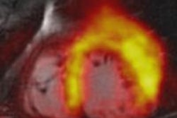
The ability of cardiac magnetic resonance (CMR) imaging to accurately measure ventricular function, myocardial scarring and ischemia is now generally accepted and reflected in guidelines, which recommend CMR alongside nuclear perfusion imaging and stress echocardiography.
 Dr. Sven Plein. All images courtesy of the European Society of Cardiology (ESC).
Potential advantages of CMR over nuclear imaging are its higher spatial resolution, the anatomical correlation provided and the fact that CMR does not expose patients to ionizing radiation.
Until recently, however, the evidence base for the use of CMR in patients with coronary artery disease (CAD) has been sparse, and CMR practice was limited to a small number of specialist centres. Both of these limitations are being overcome.
Two recent studies have compared CMR with SPECT in larger populations. The single-center CE-MARC study showed in 752 patients that CMR had superior sensitivity and negative predictive value to SPECT to detect angiography-defined CAD, while specificity and positive predictive value were similar.1 The multicenter MR-IMPACT2 study confirmed the superior sensitivity of CMR over SPECT in 533 patients, albeit with inferior specificity.2 Outcome and cost-effectiveness data from CE-MARC will become available next year and several further CMR outcome studies are nearing completion.
In parallel, the availability of CMR is rapidly expanding. In the U.K., for example, CMR is now performed in over 60 centers, having crossed the boundary from specialist centers to district hospitals. In Europe many CMR centers have a throughput comparable to large nuclear imaging departments. The accumulating evidence therefore is that CMR can replace nuclear imaging as a safer (no ionizing radiation) and potentially more accurate first-line test for the detection of CAD.
The transition from nuclear imaging to CMR will not and should not be abrupt, however, and will initially remain governed by local expertise, availability and, regrettably, turf-wars. At the same time, continuing technological development in both imaging modalities will provide substance for research, debate and re-evaluation. But it is now timely to invest in CMR infrastructure on a larger scale and to accelerate the training of physicians in CMR, in anticipation of a gradual transition from nuclear imaging to CMR.
References
- Greenwood JP, Maredia N, Younger JF, et al. Lancet. 2012;379:453-460.
- Schwitter J, Wacker CM, Wilke N, et al. Eur Heart J. 2012; Mar 4 [Epub ahead of print].
NO, says Dr. Erick Alexanderson Rosas, National Heart Institute Ignacio Chavez, Mexico
Nuclear cardiovascular imaging involves four well known techniques: single photon emission computed tomography (SPECT), positron emission tomography (PET), and hybrid techniques SPECT/CT, and PET/CT.
 Dr. Erick Alexanderson Rosas.
SPECT imaging conveys many important advantages in availability, spread, low cost, fast acquisitions, and an average 20-year well-consolidated experience with the method. Its diagnostic and prognostic utility has been proven by years of intensive research in clinical conditions with an overall technique accuracy of more than 90% with great interventional therapy guidance.
In Mexico there are now more than 100 SPECT centers spread throughout the country, while access to CMR imaging today relies upon roughly 10 centers.
Nuclear cardiology acquisition protocols are simpler than CMR acquisition sequences, which have far more elaborate bases.
PET scanning offers important improvements over SPECT, including better spatial resolution and reduced radiation exposure. Meta-regression demonstrated that CMR and PET have a significantly higher diagnostic accuracy than SPECT, on a patient and coronary territory basis. Moreover, PET allows the absolute quantification of myocardial blood flow in different physiological and pathological conditions (a proven and useful method), enhancing the meaning of qualitative and semiquantitative assessments; CMR offers this possibility as well, but so far as a less developed and proven approach.
Hybrid imaging has arisen as a response to the intrinsic problems of single nuclear modalities. This is how PET/CT and SPECT/CT now consider attenuation correction and the fusion modality to assess anatomical and metabolic topography of the heart. Clinical validation studies show that advances in attenuation correction lead to an increase in the specificity of SPECT, with fewer false positive interpretations.
On the downside of nuclear methods, controlled doses of radiation are inevitable, unlike CMR, which offers the advantage of radiation-free acquisitions. Nevertheless, PET radiopharmacy has recently developed ultra-short half-life tracers, which considerably reduce radiation exposure and therefore potential adverse effects.
It is true that CMR’s spatial resolution is considerably higher, allowing the detection of microsubendocardial infarctions and equally small ischemic areas of the myocardium -- but whether these areas are of clinical relevance is yet to be proved.
Editor's note: This article was originally published on 29 August in the European Society of Cardiology's (ESC) daily newspaper, ESC Congress News.


