Screening cases of craniofacial gunshot wounds using CT angiography (CTA) may help diagnose unexpected cervical vascular injuries remote from the penetrating tract, according to a major new study of 427 patients who sustained gunshot wounds to the head, face and/or neck.
The significance and optimal therapy of these injuries are unknown, and additional experience will be needed to determine the significance of "indirect" cervical arterial injuries in the setting of craniofacial gunshot wounds, noted the authors of an article published online on 9 May by European Radiology.
Cervical arterial injuries spatially remote from the wound tracts occur with a frequency comparable to that of blunt cerebrovascular injuries (BCVI) in blunt trauma populations, and may result from a similar mechanism of injury that is most likely related to sudden, violent hyperextension of the head and upper neck, explained lead author Dr. Scott D. Steenburg, from the emergency radiology section of Indiana University School of Medicine, Indianapolis, U.S. Until the clinical consequences of these indirect arterial injuries are known, radiologists who think it is important to screen patients for BVCI should consider adding craniofacial gunshot wounds as an indication for screening for cervical arterial injuries, particularly in patients with other injuries thought to be survivable, he added.
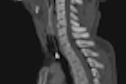
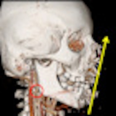
 Top: 3D volume-rendered image of a patient following a submental shotgun wound. The yellow arrow indicates the trajectory of the gunshot wound and the red arrow encircles the right internal carotid artery (ICA) pseudoaneurysm. Bottom: Sagittal oblique maximum intensity projection (MIP) image demonstrates ICA pseudoaneurysm, indicated by the yellow arrow. All images courtesy of Dr. Scott Steenburg.
"Up to 20% of patients with blunt cervical arterial injuries do not have the traditional 'high risk' factors, and up to 10% of patients have no other associated injuries," he noted in an email to AuntMinnieEurope.com. "We hope that this study will bring to light an additional risk factor for identifying patients with unsuspected cervical vascular injuries."
The literature on pure blunt cervical vascular injuries is quite clear: Diagnosing and treating asymptomatic cervical vascular injuries significantly reduces the risk of subsequent stroke, Steenburg stated. The results of this study may alter imaging protocols and practice patterns for clinicians who treat patients with craniofacial gunshot wounds, though he admits the small study population makes it difficult to draw any definitive conclusions as to the significance of this injury.
Steenburg carried out the research with Dr. Clint Sliker while they both worked at the diagnostic imaging department at the University of Maryland Medical Center and R Adams Cowley Shock Trauma Center in Baltimore, MD. They studied 427 patients who sustained gunshot wounds between 2006 and 2010. Of these cases, 222 underwent CTA, which yielded 78 vascular injuries in 56 patients. Forty-two patients had one arterial injury, 15 patients had two arterial injuries, and two patients had three arterial injuries. There were five internal carotid artery injuries remote from the wound tract. The incidence of these "indirect" cervical arterial injuries was 1.2%, or 2.8% of patients who underwent CTA.
The mean age was 31.9 (range 13-88 years), and 388 patients were male (90.9%). The mortality rate among the 427 patients was 49.6%. The highest mortality rate (91.9%) occurred in the subset of patients who did not undergo imaging, while the mortality in those who were imaged with unenhanced CT was 57.3%. The mortality rate in patients who were imaged with CTA was 23.4%. The equipment used included the Brilliance 16 Power, Brilliance 40, and Brilliance 64 systems (Philips Healthcare).
A limitation of the study was the retrospective design that precluded the use of a uniform imaging protocol, the authors wrote. The decision to undergo imaging, and to what extent, was at the discretion of the attending trauma surgeon.
"These findings suggest a bias of referring physicians towards excluding patients with perceived lethal injuries from further unnecessary imaging evaluation or, alternatively, a bias towards the use of CTA patients with perceived survivable injuries," they noted. "Either way, this suggests that the patient population constituting our primary study population was not homogeneous, which may have skewed the results away from the true incidence of indirect cervical arterial injuries."
The authors have no formal plans to continue this study, though they are always looking for associated risk factors for blunt cervical vascular injuries.