A new review of recent findings in MRI of patients with standard cardiac pacemakers (PMs) provides striking evidence that suggests careful use of MRI in such patients is largely event-free, but the authors warn radiologists to follow a set of best practice guidelines to reduce the risk of life-threatening events.
MRI in patients with nonMRI-compatible PMs is still far from a routine examination of slightly increased complexity, according to the authors, who say they cannot conclude that the procedure is generally safe.
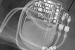
The potential hazards of MRI include the interaction of the devices and sensors with the magnetic fields that might disrupt the cardiac rhythm, and the build-up of energy in the electrode leads that can discharge heat into the tissues. MRI is, however, occasionally indicated in seriously ill patients: The likelihood that MRI will be indicated at least once in a patient with a PM is estimated at around 50%, according to the review published in the latest issue of Deutsches ärzteblatt International (May 2012, Vol. 109:15, pp. 270-275).
Provided appropriate steps are taken to optimize the procedure, the benefits of MRI will outweigh the risks, noted lead author Dr. Henning Bovenschulte, from the department of radiological diagnostics, University Hospital of Cologne in Germany. MRI should only be performed on a PM or implantable cardiac defibrillator (ICD) patient when there is no alternative such as CT, ultrasound, or nuclear medicine, state the authors, who based their recommendations on a number of published studies involving MRI in 1,043 patients with a PM or ICD.
Close cooperation between the physician who establishes the indication for MRI, the cardiologist, and the radiologist is indispensable. In each case, cardiological assessment of PM dependence or nondependence must be obtained before the examination, and if a patient is PM dependent, doctors should consider re-evaluating the need for MRI, according to the authors.
For some conditions such as intraspinal cerebral processes of degenerative, inflammatory, or neoplastic origin, with existing or threatened neurological symptoms, MRI is the sole indication and can point to the appropriate treatment. For such cases, structured multidisciplinary management at well-equipped centers with experienced staff and available emergency personnel remains the key to safety and both a radiologist and a cardiologist should continually monitor the procedure.
Good planning is indispensible. Specifically, MRI should not be performed any earlier than four to eight weeks after device implantation. Radiologists need to restrict field strength to below 1.5-tesla, keep specific absorption rate (SAR) as low as feasible, and examinations should contain as few sequences as possible -- preferably without the use of surface coils.
In addition the device needs to be inspected before and after MRI, the PM sensing and pacing settings adjusted appropriately before the exam, with pacemaker and defibrillator functions deactivated where possible, or the asynchronous pacing mode activated. Post MRI, the patient must be monitored until completion of the reprogramming and testing of the device. ICD and PM parameters such as the pacing threshold need to be checked after one month and three months. Where these precautions were observed, no life-threatening incidents occurred across the studies. In 11 cases electrical resetting of the device was necessary, and a significant increase in pacing threshold was seen in 16 cases.
Classification of patients according risk involved in performing MRI when no other diagnostic procedure is indicated
|
|||||||||
| Source: Bovenschulte H, Schlüter-Brust K, Liebig T, et al. MRI in patients with pacemakers -- overview and procedural management. Dtsch Arztebl Int 2012;109(15):270-275. |