CT is vital for evaluating the central airway, mediastinal structures, and lung parenchyma, plus it's more sensitive than plain radiographs in detection of structural changes within the lungs, but CT examinations must be clinically justified by the referring clinician and radiologist.
That's the central message of a comprehensive overview of pediatric chest CT conducted by authors from one of Europe's leading hospitals for children.
"Multidetector CT (MDCT), with its isotropic resolution and fast imaging acquisition times, reduces the need for invasive diagnostic investigations. However, users must be vigilant in their imaging techniques to minimize radiation burden, whilst maintaining good image quality," noted lead author Carolyn Young, a radiographer from the Cardiothoracic Unit at Great Ormond Street Hospital for Children NHS Trust in London.

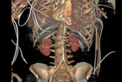
 Prospective ECG-gated study on an unsedated free-breathing 2-month-old girl with an average heart rate of 122 beats per minute (bpm). The right pulmonary artery is hypoplastic (top left). An aortopulmonary collateral arising from the celiac axis supplies the right lung, as shown in the volume-rendered image (top right) and the maximum intensity projection image (bottom). All figures courtesy of Carolyn Young.
MDCT using a low radiation dose protocol is the best diagnostic tool for assessing the central airway, cardiovascular and mediastinal abnormalities, and the lung parenchyma in children, and the relevant information can be acquired with a single-volumetric data set acquisition, she stated. Using thin-slice collimation acquisition with inherent isotropic resolution, the image data can be manipulated and reformatted to display 2D and 3D images with the same spatial resolution as the axial images, thus enhancing diagnostic accuracy and providing data that can be used in presurgical planning and patient management.
Furthermore, wider coverage with faster tube rotation speeds has reduced overall imaging time, leading to a decrease in sedation needs, improved image quality due to reduction in motion and respiratory artifacts, and high-quality depiction of small vessels due to improved contrast enhancement as a result of higher contrast concentration over a shorter time period.
"Taking all of this into consideration, imaging with MDCT is a relatively noninvasive procedure compared to conventional angiography, but it also carries a higher radiation burden," wrote Young in an online article published by Insights into Imaging on 27 March. "The use of ionizing radiation is always of concern in pediatric CT imaging, and a careful risk-benefit analysis should be done before performing CT."

 Retrospective ECG-gated study on an 8 month old with pulmonary hypertension shows narrowing of the inferior left pulmonary vein (arrow, left). The peripheral venous vessels (right) are also ecstatic, reflecting a more widespread pulmonary venous abnormality.
A dedicated bodyweight or bodysize-based imaging protocol should be developed in conjunction with manufacturers' recommendations, and the radiologist must optimize scanning parameters and tailor the CT protocol to each patient's needs, she added. The use of 120 kVp is no longer advocated in pediatric chest imaging, and should be reduced to 80 or 100 kVp without detriment to image quality.
Likewise, tube current can also be reduced, but with a resultant increase in image noise. The degree of tube current reduction depends on the level of image noise that is deemed acceptable by the reporting radiologist without degradation to the spatial resolution, thus ensuring the CT images can be accurately interpreted and a diagnosis made, according to Young. A constant image noise level can be maintained to follow the patient's anatomy by employing automatic tube current modulation (ATCM), but the tube current will increase, especially over the shoulder and at the lung base/diaphragm regions, so it is essential to limit coverage to the area of interest. Use of ATCM is debatable in smaller children as they are more rounded in body shape, making the use of angular tube current modulation ineffective.
Other possible radiation dose reduction strategies include the use of filters that are automatically deployed in spiral acquisition during the compulsory "run-up" and "run-down" period as a means of reducing the over-ranging effect that is inherent in MDCT. Adaptive statistical iterative reconstruction can reduce image noise, thus allowing the reduction of imaging parameters used. Additionally, radiation exposure modulation over sensitive organs reduces tube output when the x-ray tube is in the AP position over the breast and thyroid region, and does not require bismuth shielding.
"From our experience, children over 3 years of age are usually compliant for their procedure provided they are properly prepared through play therapy beforehand, using a mock-up toy of the scanner to take the child and their parents through the scanning process," Young noted. "It is also a good opportunity to assess the child's ability to respond to breath-holding instructions, otherwise gentle respiration is encouraged.

 Retrospective ECG-gated study on a 5 year old with severe pulmonary hypertension. The CT images show dilated pulmonary arteries (top left). There is right atrial and ventricular dilatation with hypertrophy and bowing of the interventricular septum into the left ventricle (arrow, top right). There is abnormal arborization and angulation of the peripheral pulmonary arteries (top right, bottom), with multiple peripheral centrilobular nodules with perilesional ground-glass attenuation. Features are in keeping with pulmonary capillary hemangiomatosis.
At Great Ormond Street, children less than 15 kg in weight are sedated, and general anesthetic is reserved for noncompliant patients. The nurse-led sedation team will administer a short and light-acting sedative of chloral hydrate at a dose of 50 mg/kg. The use of a more quick-acting sedative of propofol administered by an anesthesiologist has been reported with low incidence of adverse events, and the use of ketamine and midazolam has also been reported as safe and effective, she wrote.
Children should always have their intravenous access for contrast enhancement sited prior to arrival in the radiology department in order to help disassociate the CT department from the trauma of having the cannula sited, recommended Young.
Overall, MDCT has extended CT applications for use in pediatric chest imaging. It remains the gold standard for imaging in certain pathologies and is especially helpful when a diagnosis can be reached without the need for more invasive diagnostic investigations. Radiation burden remains a major concern, and users must be vigilant in their dose reduction techniques, while maintaining good image quality which is "fit for purpose," she concluded.

 A 14-year-old patient with transposition of greater arteries and gross pulmonary arterial hypertension. CT angiography study shows aneurysmal dilation of the pulmonary arteries (top left, top right), with thrombus in the right middle lobe pulmonary artery (bottom left). There is partial anomalous pulmonary venous return from the right upper lobe to the superior vena cava (arrow, bottom right).

 Same 14-year-old patient, there is mark dilatation of the pulmonary tree, causing compression of the trachea at the carina (top) and on the left main bronchus behind the aneurysmal right pulmonary artery (bottom)