Speckle-tracking echocardiography (STE) is increasingly regarded as a viable and cost-effective means to measure infarct size, noted presenters at the European Association of Echocardiography's annual congress, Euroecho & Other Imaging Modalities, held last week in Budapest.
Infarct size quantification is typically not measured in routine clinical practice, because the reference method of an MRI examination using gadolinium-based contrast agents is expensive and time-intensive. But STE can be carried out by cardiologists using portable equipment at a patient's bedside at a cost that is significantly less expensive than a MRI with contrast examination.
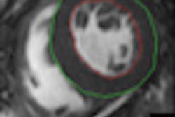
STE is a noninvasive echocardiography technique that works by tracking the movement of natural acoustic markers, or "speckles," which are visible on monochrome ultrasound images. With the use of wall motion tracking software, the speckle movement, and therefore myocardial tissue movement, can be visualized during the cardiac cycle.
 The Euroecho & other Imaging Modalities meeting was organized by the European Society of Cardiology and included a bustling technical exhibition. Image courtesy of Robin Marshall.
Speckle tracking can be used to evaluate myocardial strain, which describes the myocardial deformation throughout the cardiac cycle. Reductions in measurement of strain have been found to show direct relationships to the size of an infarct.
Infarct size matters for determining how well patients will recover from a ST segment elevation myocardial infarction (STEMI), the complete blockage of a coronary artery by a blood clot. Statistics suggest that people who suffer damage to more than 30% of the left ventricle are twice as likely to die within a year of the event than people who suffer less damage.
European Association of Echocardiography (EAE) President Dr. Luigi Badano of the University of Padua in Italy told congress attendees that evidence is mounting that screening for patients with larger infarct sizes enables identification of patients with a worse prognosis who benefit from more aggressive therapy and more frequent follow-up visits. "It's well known that patients with larger infarcts are more likely to undergo alterations in the dimensions, mass, and shape of the left ventricle. Such cardiac remodeling leads to heart failure. Many more options exist for STEMI patients deemed at high risk of adverse events," he said.
Studies presented
Results of studies of the use of 2D and 3D speckle tracking were presented in scientific sessions at the meeting.
At the University of Padua, Dr. Denisa Muraru and colleagues used 3D speckle tracking to estimate infarct size and necrosis transmurality in patients with recent STEMI who had undergone successful coronary angioplasty treatment.
The researchers assessed 49 patients were assessed by 3D speckle tracking, and compared the obtained left ventricle strain parameters with peak troponin I levels, as an estimate of the extent of myocardial cell injury. In a subgroup of 27 patients who underwent additional assessment with a delayed enhancement MRI exam within 24 hours from the echocardiographic study, circumferential strain showed the closest correlation with infarct size and the best predictive power to identify left ventricle segments with transmural necrosis among all strain components.
"Our preliminary study demonstrates that 3D circumferential strain could be used as an accurate and reproducible marker for infarct size estimation by ultrasound in STEMI patients," she said. "Long-term follow-up will be needed to verify if 3D strain parameters improve the predictive prognostic value of conventional parameters after STEMI."
One of the advantages of 3D over 2D speckle tracking cited by Muraru is that it allows the assessment of longitudinal, circumferential, radial strain, and area strain at the same time.
A Bulgarian study presented by Dr. Krasimira Hristova and colleagues from the University National Heart Hospital in Sofia investigated the ability of 2D speckle-tracking echocardiography using a vector velocity imaging technique to determine infarct size.
In this study, 30 patients who had a coronary angioplasty for an acute myocardial infarction within 24 hours, and a control cohort of 20 healthy volunteers were assessed with both vector velocity speckle tracking and intracoronary electrocardiography. The results showed that in patients who had suffered STEMI radial and circumferential strain decreased in the infarct area, preinfarct area, and remote regions acutely in comparison with the control group. However, longitudinal strain was only decreased in the actual infarct area, Hristova reported.
"While longitudinal strain shows the best relationship to infarct size, we believe that radial and circumferential strain may be useful to predict the later development of adverse left ventricle remodeling," she said. Hristova stated that in the next segment of the 2D speckle tracking study, she and her colleagues hope to be able to compare their initial results for longitudinal, radial, and circumferential strain with the longer-term effects on left ventricular remodeling.