For digital mammography, using local contrast optimization for image processing may increase perceived suspiciousness of breast tissue, but diagnostic accuracy may remain similar to other algorithms, Dutch researchers found in a study published online on 10 November in European Radiology.
Digital mammography requires image processing before images are suitable for display, but there is no standard algorithm and optimization varies. "Because of the lack of easy and objective methods for measuring processed image quality, however, we often have to rely on the impression experts have of the appearance of images to rate image processing," noted lead author Dr. Roelant Visser, from the National Expert and Training Centre for Breast Cancer Screening in Nijmegen, the Netherlands, and colleagues.
Current image processing algorithms aim for a maximum local contrast while decreasing the total image dynamic range, they wrote. The contrast optimization techniques can have a large effect on the appearance of images -- affecting diagnostic accuracy -- and could influence the perceived suspiciousness of healthy breast tissue.
However, in their study, the researchers found no significant difference in diagnostic accuracy of the processing algorithms they analyzed by comparing the areas under the receiver operating characteristic curves. Visser and colleagues collected data from a screening region in the Netherlands consisting of 263 digital screening cases (153 recalled, 110 normal). Each case was available twice, once processed with a tissue equalization algorithm and once with local contrast optimization. All cases had digitized previous mammograms. For both algorithms, the probability of malignancy of each finding was scored independently by six screening radiologists.
     |
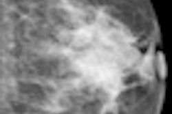
| Example of a subtle finding in a right-sided mediolateral oblique view, which was only reported by radiologists when using premium view (PV). A: Digitized prior. B: Tissue equalization (TE) processed image. C: PV processed image with the annotation. D: The resulting image of subtracting TE from PV. E: Thresholded version of D. White areas indicate that pixels in the PV image have relatively higher intensity than the related pixels in the TE image, whereas black areas indicate the opposite. In PV images, low-frequency trends are suppressed (no noticeable signal decrease in the breast edge in PV compared with TE), whereas higher-frequency structures are emphasized (e.g., glandular structures). All images courtesy of Wouter Veldkamp, PhD, Leiden University Medical Center. |
The cases were acquired using the Senographe Essential digital mammography system (GE Healthcare). Tissue equalization is a standard GE application that corrects for low-frequency variations resulting from under- and overpenetration of x-rays. As a result, the image dynamic range is reduced, enabling improved soft-copy image display.
The local contrast optimization, premium view, has been designed to improve the quality of the information presented to the radiologist for diagnosis and also the reading speed by optimizing the local contrast in breast structures. In premium view, low-frequency structures are obtained from the original image by low-pass filtering. High-frequency structures are obtained by subtracting the low-pass filtered image from the original image. The low- and high-frequency images are both processed and weighted individually, then added together. The resulting image exhibits reduced contrast between different tissue types but enhanced contrast of small-scale anatomical architecture.


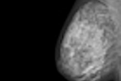
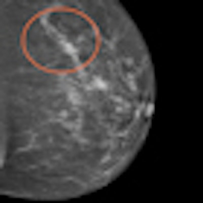
 Another example of a finding in a left-sided craniocaudal view that was reported clearly more often by radiologists when using premium view (PV). A: Tissue equalization (TE) processed image. B: PV processed image with the annotation. C: Similar to image above, the resulting image of subtracting TE from PV. D: The thresholded version of C.
For all six radiologists, perceived case suspiciousness -- defined as the highest probability of malignancy of all radiologist findings -- was higher using premium view optimization.
The major difference between the processing algorithms was an additional local contrast optimization when premium view was applied. "Premium view is aimed at increasing the visibility and suspiciousness of malignant lesions, but in our study the perceived suspiciousness of benign lesions and normal cases is increased as well," the researchers wrote. "An effect of local contrast enhancement could be that both normal (dense) structures and abnormal structures appear more suspicious due to their enhanced signal."
High-contrast images are usually preferred because of the increased visibility of the lesions, but the algorithm also affects normal cases. "In our study, the perceived suspiciousness of the normal cases increased even more than that of the malignant cases," they noted.
Future studies should investigate the influence of both the learning effect and the degree of similarity with previous mammograms on diagnosis with respect to the introduction of new postprocessing methods, the researchers advised.
"This study examines just two out of many possible combinations of appearances of currents and previous mammograms," they wrote. "For manufacturers of digital mammography systems, image appearance has become an important means of distinguishing themselves from each other. Previous studies have suggested that algorithms using contrast enhancement techniques may improve diagnostic accuracy. This effect is not convincingly present in our study. Our study suggests that the introduction of new image processing algorithms is likely to influence the recall rate because of changes in perceived case suspiciousness while diagnostic accuracy may be similar."
The researchers will continue to evaluate postprocessing algorithms in observer studies with radiologists, said study author Wouter Veldkamp, PhD, in an interview with AuntMinnieEurope.com. "This should contribute to a better understanding of the relationships between the observer (radiological) outcome and the physical characteristics of the images."
"We expect our research line to generate new knowledge that can be used to optimize currently available image postprocessing algorithms," he said. "Furthermore, we are interested in developing objective quality tests to quantitatively evaluate image quality after postprocessing."