Validated imaging tools need to be incorporated into patient care to enable a shift from the current paradigm of simply identifying the presence or absence of a tumor and assessing cytotoxicity. Furthermore, the shift toward a future paradigm of biological characterization of both the tumor and the individual, and using these tools to stratify patient response to therapy, are vital to assist the development of personalized medicine.
That's the view of Dr. Sandy McEwan, chair of the department of oncology at the University of Alberta in Edmonton, Canada, who gave the Marie Curie lecture at the European Association of Nuclear Medicine (EANM) annual congress, held this week in Birmingham, U.K.
"Imaging must come to help develop an understanding of tumor biology, not just tumor structure," McEwan said. He added that the molecular imaging of cancer requires appropriate use and consolidation of many technologies already available, and characterizing the internal environment of a tumor using imaging biomarkers will also allow a better estimation of prognosis and outcome, as well as predicting an assay of treatment response.
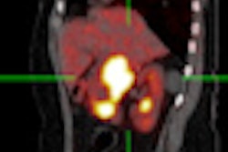
 The impending shutdown of the Chalk River reactor in Canada has initiated research into alternative methods of production of technetium-99m, particularly using small medical cyclotrons. These images are the first GMP/GCP images of biodistribution of Tc-99m pertechnetate made on a cyclotron (C-Pert) compared with generator produced Tc-99m pertechnetate (G-Pert). This study is a collaboration among the University of Alberta, the University of Sherbrooke, and ACSI. All images courtesy of Dr. Sandy McEwan.
It is clear from CT images that there are visible, morphological differences within the boundary of a single tumor, be it primary or metastatic, shown by differences in enhancement, he explained. These differences represent the intricately complex microenvironment and infrastructure, and the various biological aspects of an individual tumor.
On the one hand, techniques for oncologic imaging that only assess changes in the measurement of tumor size and volume, as well as levels of cytotoxicity, can be of limited value. On the other, many emerging therapies, such as cytostatic agents, target tumor growth and metabolism while having little effect on tumor mass for up to nine months.
Based on current imaging and clinical trials criteria, these therapies are considered failures, yet such criteria do not adequately evaluate the newest generation of anticancer therapies, according to McEwan. He advocates that a move from the current, 'reductionist' view of imaging in cancer is essential if there is to be a move toward enhancing personalized care in oncology and radiology.
Acquired capabilities of cancerous cells, such as the potential to evade apoptosis, support sustained angiogenesis, and divide limitlessly, can be assessed using various imaging methods. PET and MR tracers exist to evaluate, for example, the cellular energetics of a tumor, cell death, and proliferative signaling, but there has been no framework put in place to incorporate such vitally important clinical tools into overall cancer care, he commented.
 FDG and FES images in a patient with breast cancer metastatic to bone. The FES image shows heterogeneity of receptor expression at sites of bone metastases defined by FDG. This suggests the possibility of using FES as a means of stratifying patients for most appropriate treatment.
For images to be used as a potentially true molecular biomarker by oncologists and radiologists alike, clinical trials challenges must be addressed by "linking imaging to 'omics,' " McEwan pointed out. Such linkages do exist, as in the case of PET and MR tracers, but they need to be developed upon and integrated into a standardized clinical capacity. To do this, the necessary clinical trials capability must be put in place, and there must be development of the translational pipeline to integrate imaging biomarkers into drug discovery through multicenter trials, combined with standardization of chemotherapeutic protocols and data harmonization.
In his appeal for a paradigm shift in the molecular imaging of cancer, toward biological characterization of tumors and away from tumor structure, McEwan stressed the importance of appropriate training of young physicians and scientists in molecular biology and using imaging biomarkers. Using imaging tools to recognize and understand the different biological aspects in the microenvironment of an individual tumor is the first step in developing a personalized approach to cancer care. The image will only become a true molecular biomarker with multicenter, multinational collaboration and cross-center validation, he stressed.
For McEwan, understanding the relevant biological characteristics underlying an individual's disease and adapting patient care accordingly must be the ultimate objective of personalized medicine. Molecular imaging plays a crucial role in obtaining such highly specific, personalized diagnoses and treatments, but the acquired capabilities and microbiology of tumors make this personalized visualization and characterization of disease all the more difficult. So, too, does the current paradigm of molecular imaging in oncology, he concluded.
 The FLT image is of a patient with non-small cell lung cancer (NSCLC) showing marked positive uptake. Marechal has shown that survival following nucleoside-based chemotherapy correlates directly with the expression of human equilibrative transporters. Paproski has shown that gemcitabine and FLT have the same transport into cells in vitro. It is possible to hypothesize that FLT uptake in a patient with NSCLC will be a predictor of response to nucleoside-based chemotherapy.