A European Union-funded multicenter collaborative project is intensifying its efforts to develop objective and efficient methods for enabling earlier diagnosis of Alzheimer's disease.
PredictAD aims to extract biomarkers from heterogeneous patient data and integrate them for objective and evidence-based diagnostics. For example its reasearch team is exploring how MRI can be used to measure atrophy in the mediotemporal lobe, which is recognized as a hallmark of Alzheimer's.
In current clinical practice, images are interpreted mostly only by visual inspection, but there is a need for objective measurements, according to Dr. Jyrki Lötjönen, the scientific coordinator of the project and principal scientist and adjunct professor of signal and image processing at the VTT Technical Research Centre in Tampere, Finland.
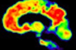
![Upper row shows MR images from a healthy person and a patient with Alzheimer's disease (AD). The right image shows clear signs of atrophy. The lower row shows PET images acquired with the amyloid imaging agent [F18]flutemetamol. The left image shows low uptake in the cortical regions, indicating no presence of amyloid plaques, while the right image shows high uptake in the cortical regions. The information from these modalities is complementary; the PET image shows information related to the underlying AD pathology, whereas the MR image shows information related to neuronal loss, a downstream effect of the disease. [F18]flutemetamol is a compound under development, and the data are from the Phase II trial). Image courtesy of Lennart Thurfjell, PhD.](https://img.auntminnieeurope.com/files/base/smg/all/image/2011/06/ame.2011_06_17_08_36_33_774_2011_06_17_predictAD_scans.png?auto=format%2Ccompress&dpr=2&fit=max&q=70&w=700) Upper row shows MR images from a healthy person and a patient with Alzheimer's disease (AD). The right image shows clear signs of atrophy. The lower row shows PET images acquired with the amyloid imaging agent [F18]flutemetamol. The left image shows low uptake in the cortical regions, indicating no presence of amyloid plaques, while the right image shows high uptake in the cortical regions. The information from these modalities is complementary; the PET image shows information related to the underlying AD pathology, whereas the MR image shows information related to neuronal loss, a downstream effect of the disease. [F18]flutemetamol is a compound under development, and the data are from the Phase II trial). Image courtesy of Lennart Thurfjell, PhD.
Upper row shows MR images from a healthy person and a patient with Alzheimer's disease (AD). The right image shows clear signs of atrophy. The lower row shows PET images acquired with the amyloid imaging agent [F18]flutemetamol. The left image shows low uptake in the cortical regions, indicating no presence of amyloid plaques, while the right image shows high uptake in the cortical regions. The information from these modalities is complementary; the PET image shows information related to the underlying AD pathology, whereas the MR image shows information related to neuronal loss, a downstream effect of the disease. [F18]flutemetamol is a compound under development, and the data are from the Phase II trial). Image courtesy of Lennart Thurfjell, PhD.To meet this need, the PredictAD team has developed tools for measuring the size of the hippocampus, the atrophy rate of the hippocampus, and two approaches based on comparing patient data with previously diagnosed cases available in large databases. PET is also being studied in the project. A novel tracer developed recently especially for diagnostics of Alzheimer's disease provides promises for very early diagnosis of the disease.
Diagnosis requires a holistic view of the patient that combines information from several sources, such as clinical tests, imaging, and blood samples, Lötjönen explained.
"The aim of the PredictAD project is to develop an objective indicator to diagnose Alzheimer's disease at the earliest stage possible," he said. "Current diagnostic guidelines emphasise the importance of various biomarkers in diagnostics. We have developed novel approaches to extract biomarkers from imaging data, electrophysiological data and blood samples, and a unique and clinically useful software tool for integrating all these heterogeneous measurements."
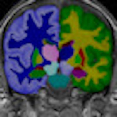
 Segmentation of brain images is often a prerequisite for quantitative analysis of images. Image courtesy of Dr. Jyrki Lötjönen.
Alzheimer's disease is known to affect the electromagnetic activity of the brain. The PredictAD group is studying the performance of transcranial magnetic stimulation (TMS) combined with electroencephalographic (EEG) measures in detecting the disease. The strength of TMS/EEG is that it allows direct and noninvasive perturbation of the human cerebral cortex without requiring the subject's collaboration, according to Lötjönen. His team's research has shown significant changes in Alzheimer's patients compared with healthy aging people.
Molecular level biomarkers are currently under extensive investigation in Alzheimer's research, he explained. Many biomarkers, such as tau proteins and b-amyloid 42, measured from the cerebrospinal fluid (CSF), have been found to be strongly related with the disease.
A major challenge of these biomarkers is that taking samples from CSF is an invasive measurement limiting their usability in early diagnostics. Blood samples would be an excellent source for detecting Alzheimer's disease as blood sampling is not considered an invasive technique, Lötjönen said. PredictAD has studied the role of metabolomic and protein compounds in Alzheimer's disease from blood samples, and the preliminary results reveal several promising compounds.

 Left: Regions showing severe atrophy for an Alzheimer's patient during a 24-month follow-up period. Right: The size of the hippocampus is a well-known biomarker in Alzheimer's disease. The green contours show the automatic segmentation result of the hippocampi. Image courtesy of Dr. Jyrki Lötjönen.
Currently, clinicians make the final diagnosis by combining heterogeneous measurements with information from interviews of the patient and relatives. This process involves subjective reasoning and requires strong expertise from the clinicians. Modern hospitals have huge data reserves containing hidden information that could be utilized in diagnostics by systematic mathematical modeling, Lötjönen noted.
According to Dr. Gunhild Waldemar from Copenhagen University Hospital in Rigshospitalet, "Successful, early diagnostics combined with the novel drugs under development and early psychosocial care may delay the institutionalization of patients, reducing suffering and the costs to the society. It has been calculated that delaying the onset of the disease by five years would halve all costs of Alzheimer's disease, and delaying onset and progression by only one year would reduce the number of Alzheimer's cases by about 10%."
Companies like GE Healthcare, and pharmaceutical manufacturers, are investing heavily in this area, and commercialization of the results is already ongoing in PredictAD, noted Lennart Thurfjell, PhD, head of diagnostic software for medical diagnostics at GE Healthcare in Uppsala, Sweden, who is leading the activities of dissemination and exploitation.
 The objective of the PredictAD project is to extract biomarkers from heterogeneous patient data and integrate them for objective and evidence-based diagnostics. Image courtesy of Dr. Jyrki Lötjönen.
To present and discuss the results of the PredictAD project and recent innovations for the early diagnosis of Alzheimer's disease, PredictAD organized a symposium in Kuopio, Finland, on 15 June 2011. The event, "From Patient Data to Personalized Healthcare in Alzheimer's Disease," was designed to coincide with the Kuopio Bio-NMR Workshop: MRI of stroke, epilepsy and neurodegenerative diseases.
The Kuopio Bio-NMR Workshop aimed to bring together aimed to bring together international MRI researchers from both experimental and clinical research environment, and was for graduate students, post docs, and more advanced research personnel, as well as medical doctors who are interested in applying MRI in their research or work. The workshop covered topics from basic principles of MRI techniques to applications in studies of neurodegenerative diseases and finally to clinical studies. The first day was dedicated to MRI techniques, and the second day to applications in experimental animal models or in clinical settings.
The following eight research, academic, industrial and medical organizations from five different European countries are involved in the PredictAD project: VTT (Finland), GE Healthcare (U.K.), Nextim (Finland), University of Eastern Finland (Finland), Imperial College London (U.K.), Karolinska Institutet (Sweden), University of Milan (Italy), and Copenhagen University Hospital, Rigshospitalet (Denmark).