For the first time in its 92-year history, the German Congress of Radiology (Deutscher Röntgenkongress, DRK) took place in Hamburg. After residing in Berlin for many years, the DRK moved from Berlin to Hamburg, the second largest city in the home country of radiology. As an attendee of past meetings, I cannot say that Berlin was bad at all. Yet, definitely, it is always fascinating to explore a previously unknown congress location and to get inspired by its new set-up, architecture, and urban surrounding.
In all of this, Hamburg was a wonderful host for this 6th annual meeting hosted by both the Austrian and the German radiological societies. It all started and ended with the excellent weather, temperatures of up to 30° C during the whole meeting from 1-4 June. And due to the absence of rain, wonderful sunsets at the nearby "Alster" could be enjoyed in lakeside restaurants and bars together with national and international colleagues to discuss the hottest topic of the past days. And there were many, as the program covered a broad spectrum, starting with numerous refresher courses, new commercial products presented by the industry in the large technical exposition area, and finally the scientific sessions as the academic core of the meeting.
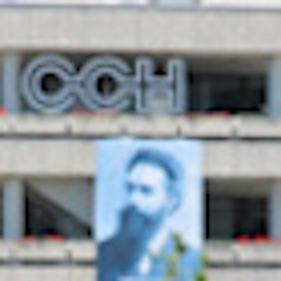
 The Congress Center Hamburg (CCH) is located next to the Planten un Blomen, a green and quiet inner-city park ideal for networking and relaxing during breaks between the sessions of this oldest meeting dedicated to medical imaging. All images courtesy of Dr. Matthias Dietzel.
It was not only the city center of Hamburg, but also the congress location itself that was ideal for networking and exchanging the latest news in radiology. Opened in 1973, the Congress Center Hamburg (CCH) has hosted many large medical congresses before. The CCH seemed to be ideal, as it is located in the very heart of the city with the historic Dammtor railway station within walking distance. So it was easy to reach, by long-distance trains, suburban trains, buses, or even by foot. Due to excellent organization, it was easy to find the way between the technical exposition and registration area to the different session rooms. As in previous years, such were named by famous pioneers of radiology -- e.g., Curie, Hounsfield, and of course Röntgen himself.
The program
For the first time in its nearly 100 years of history, the congress presidents chose a motto for the meeting: "Radiology is diversity" ("Radiologie ist Vielfalt"). And indeed it is true this specialty is amongst the medical professions covering the broadest spectrum of diseases and pathologies. Accordingly, this year's congress presidents Dr. Bernd Hamm and Dr. Walter Hruby aimed to cover both the basic current knowledge in radiology as well as highly interesting innovations and new approaches for the future. The program was split into two main components: the educational and scientific programs.
Educational program
480 refresher courses covered basic knowledge in radiology. Due to this abundance, the attendees had to choose between such courses. As in the past years, one of the most popular sessions within the educational program was the FFF-series ("Fit für den Facharzt": "Fit for the board certification examination"). It is intended for radiology residents preparing for this challenging examination. It was highly popular and it was not always easy to get good seats! Yet, such were essential to answer the excellent case-based questions with the TED-System. Presenters were all well-known specialists.
Another highlight was the refresher courses by the experts in chest imaging. During the minicourse High Resolution Computed Tomography (HR-CT), the speakers explained the importance of both structured analysis and structured reports in this challenging field of medical imaging. Starting with the "secondary lobulus," Dr. Okka Hamer gave the audience an excellent approach to HR-CT and how it should be read to get an accurate differential diagnosis of nodular lesions. Further pathologies, e.g., reticular changes, consolidations, and ground-glass opacities, were covered by Dr. Katharina Marten-Engelke and her husband Dr. Christoph Engelke.
Of course it is not possible to summarize all the excellent courses of the educational program. Yet, one entirely new course during the congress needs to be mentioned. The German Radiological Society organized an interventional hands-on workshop at the "European Surgical Institute." The latter is a unique place to train in procedures of interventional vascular radiology. For this purpose it provides excellent tutors and simulation equipment.
"As this institute is only a half-an-hour bus ride from the CCH, we had the idea to provide the attendees of the congress this highly innovative course ... where interventional novices are trained for three hours by experts in this field," Dr. Walter Gross-Fengels said. All 30 attendees lucky enough to get one of the few tickets for this workshop were enthusiastic about this great teaching.
 Residents took a break to unwind at the end of a long, hard day's work. Young Investigator Sessions included highly innovative scientific projects and were presented by young academic radiologists. They covered both molecular imaging and clinical research.
Scientific program
Altogether a total of 400 scientific presentations were given during the four congress days. Following the motto of this year's convention, a wide range of topics was covered.
Dr. Seyfer from Marburg presented highly interesting results of her molecular imaging project. She addressed the clinically challenging differentiation between inflammations and tumor tissue. Using an animal model, she demonstrated that ultrasmall superparamagnetic iron oxide leads to a signal loss in T2* images in inflammatory tissue due to accumulation in macrophages. This effect was not seen in vital tumor tissue.
Yet, evaluating new contrast is not the only way to optimize diagnosis and to enhance accuracy. As stated in a notable talk by Andreas Dietzel, PhD, from Tübingen, bioinformatics might also contribute to this task. As the number of different tissue contrasts and images is increasing with advancing technology, the final, usually binary (i.e., benign or malignant), diagnosis gets increasingly difficult for the radiologist. Computers might help to solve this problem, yet operation of such algorithms is difficult and requires a high level of training and a systematic approach. Accordingly, the author developed software to automatically process and analyze multidimensional radiological data based on numerous computer algorithms.
"The results are promising, as only few manual steps were necessary and the results were fairly accurate," Dietzel said.
Radiology is not only diagnostic but more and more also a therapeutic speciality. Dr. Philipp Paprottka investigated the utility of radioembolization of patients with intrahepatic unresectable cholangiocellular cancer (CCC). At this stage, the current treatment of choice is systematic chemotherapy. Radioembolization on the other hand is frequently used in hepatocellular cancer and liver metastases. Accordingly, Dr. Paprottka evaluated this therapeutic approach in 33 patients with advanced CCC. Following his initial results, radioembolization was an effective and safe therapeutic option for patients with CCC.
In terms of clinical MR research, one highlight was the introduction of chemical exchange saturation transfer (CEST) imaging to breast MRI. This technique might become a new contrast for tissue characterization as it allows molecular characterization with much higher spatial resolution compared with MR spectroscopy. Also clinical data are still preliminary. Dr. Heinz-Peter Schlemmer from Heidelberg, a well-know radiologist and physicist, assumed that the basis of the contrast might be choline compounds. This allowed CEST imaging with high scan quality and excellent tissue contrast of breast tissue, he concluded in his talk.
Another technical focus was the evaluation of capillary perfusion and extracellular diffusion, using diffusion-weighted MRI (DWI-MRI). Dr. Pascal Baltzer evaluated this technique to differentiate 95 liver lesions. According to this approach, diagnosis of metastases with high diagnostic accuracy was possible in clinical practice. Dr. Rotem Lanzman evaluated this technique in a smaller collective to characterize breast lesions in MRI. According to his initial results, this method also allows accurate breast tissue characterization.
Beside such technical innovations, new clinical data on breast MRI was presented at the congress. One special focus was set by the team of Dr. Werner Kaiser, a pioneer in breast MR imaging.
They presented results demonstrating the possibility of using this technique as a potential biomarker, both to address direct end points (overall survival) and surrogates (lymph node metastases). Analyzing a large database, the authors concluded that it is particularly multivariate analysis that increases accuracy of breast MRI as a noninvasive prognostic tool.
Conclusion
After four intense congress days, there is no doubt, that the 92nd congress successfully continued the traditions of its previous meetings to bring radiologists together to discuss the latest news in their specialty. CCH was an excellent location for this venue, and Hamburg had a successful premiere as the host for this meeting. See you next year!
Dr. Matthias Dietzel is from the Institute of Diagnostic and Interventional Radiology at Friedrich-Schiller-University Jena in Germany.
The comments and observations expressed herein do not necessarily reflect the opinions of AuntMinnieEurope.com, nor should they be construed as an endorsement or admonishment of any particular vendor, analyst, industry consultant, or consulting group.
You can read more about the DRK here.