Radiologists need to be more "considerate" with their acquisition techniques, especially when patients are undergoing volumetric helical perfusion CT imaging of the thorax, abdomen, or pelvis, according to a new study to be published in the May issue of European Radiology.
High-frequency volumetric helical perfusion CT imaging potentially has a higher radiation burden than single anatomical-level acquisitions. There has been concern regarding the radiation dose patients could incur from the technique so Dr. Vicky Goh, now the chair of cancer imaging at King's College London, and colleagues sought to determine what the radiation dose was for volumetric helical perfusion CT.
They determined the radiation dose by perfusion CT was on average 1.5 times higher than that of a standard CT for the thorax, abdomen, and pelvis. The dose is not insignificant, and further efforts have to be undertaken to reduce radiation dose for the procedure to be a viable clinical technique, they concluded in their study (Eur Radiol, May 2011, Vol. 21:5, pp. 974-981).
"We need to be more considerate with our acquisition techniques -- ensuring that dose is kept as low as possible when scanning, at 80 or 100 kV, that we cover only the areas that we need to (e.g., the whole tumor) but not exceeding this," Goh said. "[We need to] consider the frequency of acquisition for the parameters we need ... and also the length of the acquisition -- hence allowing us to keep dose to the minimum necessary."
In the study, Goh and colleagues recorded dose-length product (DLP) and CT dose index (CTDIvol) in patients undergoing 4D adaptive spiral CT imaging for tumor evaluation of the thorax, abdomen, and pelvis. The study included 42 consecutive examinations. Mean DLP for the method was 1,288.8 mGy.cm. Mean CTDI was 96.2 mGy. Mean effective dose was 19.6 mSv. In comparison, mean DLP for standard CT staging for the thorax, abdomen, and pelvis was 885.2 mGy.cm. Mean effective dose was 13.3 mSV.
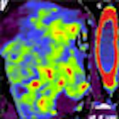
 Helical perfusion CT depicting a renal tumor acquired at 100 kV. Images show 3D image (A), B) blood flow (B), blood volume (C), and flow-extraction product (D). Images courtesy of Dr. Vicky Goh.
The mean effective dose for volumetric helical perfusion CT confirms that the helical techniques impose a much higher radiation dose than conventional radiography and CT, the authors wrote.
To put this into clinical perspective, the dose from a perfusion CT examination, while higher than CT of a single body part, is not dissimilar to some of the commonly performed CT examinations in oncology, such as multiphasic CT organ studies, in which effective doses range from 12 to 26 mSv, the authors wrote.
Since publishing the article, Mount Vernon Hospital, where Goh performed the study before transferring to King's College London, has tightened up its acquisition procedures such as length of acquisition: 45 seconds where coverage is greater than 10 cm.
"We scan at 80 kV for the neck and thorax, or 100 kV for the abdomen and pelvis, and improve[d] scan quality using postprocessing dynamic filtering techniques (e.g., 4D noise reduction)," she said.
The values recorded in the study highlight the need for clinician awareness of the radiation dose imposed by high-frequency CT acquisitions, the need to monitor dose in each patient, the need for quality control, and the need to instigate dose reduction strategies, the authors concluded.