NEW YORK (Reuters Health), Apr 30 - Pericardial adipose tissue (PAT) seen on CT scans is a risk factor for coronary atherosclerosis, according to a report in the May issue of Arteriosclerosis, Thrombosis, and Vascular Biology.
"PAT is clearly associated with coronary disease and can be considered as a risk factor," Dr. Alexander W. Leber told Reuters Health. "It may allow us to identify those patients who do not look like the typical risk patient because of a normal body habitus."
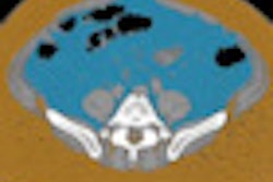
Dr. Leber from University Hospital of Munich, Germany, and colleagues simultaneously assessed pericardial fat volume and coronary atherosclerosis using dual source CT angiography in 286 patients with an intermediate pretest likelihood for coronary artery disease.
PAT volume was significantly higher in patients with any coronary plaque than in patients without coronary plaques, the authors report, and it correlated with the number of diseased coronary segments.
Increased PAT volume was also associated with lower adiponectin and HDL cholesterol levels and with higher levels of TNF-alpha and high-sensitivity CRP.
"My vision about incorporating PAT in the current risk stratification is to use it in addition to calcium scoring, for example," Dr. Leber said. "We observed that PAT accumulation precedes the development of calcified plaques, so that we are able to detect the disease in an earlier stage. I also think that PAT thickness assessment should be part of the routine echocardiogram and may be used as a surrogate marker for preclinical atherosclerosis."
"Though practical constraints (cost and radiation exposure) limit the routine use of CT scan to estimate individual cardiovascular risk at the population level, this study contributes to the fascinating discussion regarding the potentially causal role of fat abundance around the heart in coronary atherosclerosis," write Dr. Karine Clement from Universite Pierre et Marie Curie-Paris, France, and colleagues in a related editorial.
The report, the editorial concludes, "illustrates the need to investigate the additional benefit of precisely quantifying pericardial fat per se (rather than detecting it as a morphological sign of coronary alteration) as a valuable and independent coronaropathy risk factor or as a marker of visceral abdominal fat, which is known to be inflamed in metabolic disease."
By Will Boggs, M.D.
Arterioscler Thromb Vasc Biol 2009;29:781-786.
Last Updated: 2009-04-29 15:03:13 -0400 (Reuters Health)
Related Reading
PET/CT confirms presence of brown fat in adults, April 22, 2009
Copyright © 2009 Reuters Limited. All rights reserved. Republication or redistribution of Reuters content, including by framing or similar means, is expressly prohibited without the prior written consent of Reuters. Reuters shall not be liable for any errors or delays in the content, or for any actions taken in reliance thereon. Reuters and the Reuters sphere logo are registered trademarks and trademarks of the Reuters group of companies around the world.