LONDON - A final breakdown of imaging statistics from the London Olympics shows the toll that competitive sport can have on the bodies of athletes, with radiologists chronicling more than 2,300 scans acquired during the games. The breakdown was revealed last night during a U.K. Royal College of Radiologists (RCR) lecture held to mark the inaugural International Day of Radiology.
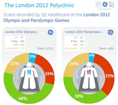
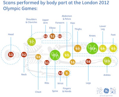
Dr. Phil O'Connor, imaging lead for the 2012 Games, said that 835 MRI, 405 x-ray, 392 ultrasound, and 79 CT examinations were performed. Of the total of 1,711, the largest amount (16.9%) was performed on knees, followed by the spine (13.2%), and ankles (12.9%). The total figures for the Paralympics were 254 MRIs, 216 x-rays, and 157 ultrasound and 28 CT scans.
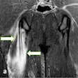
 All images courtesy of GE Healthcare.
The nature and frequency of images provided a surrogate marker tracking sporting events happening outside of the Olympic polyclinic, where the radiology service was housed, according to O'Connor.
"In fact, as the sports changed, so the injury patterns changed," said O'Connor, a consultant musculoskeletal radiologist from Leeds, U.K. "You didn't have to see any of the games anyway because you knew what was going on from the injuries coming in."
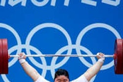
The polyclinic's radiology team saw 14 ulnar collateral injuries of the elbow. O'Connor reported that, classically, these injuries are seen in throwing events, but at the Olympics all but one was from judo and weightlifting. "We had six in one day and that was because we were reaching the finals of judo and weightlifting, but the next day it all stopped and we had lots of hamstring tears or Achilles' problems because track and field had arrived," he said.
'More pathology in 2 weeks than 10 years of practice'
O'Connor has been a specialist in sports radiology for 16 years, and ran the imaging services at the 2002 Commonwealth Games and the 2003 World Indoor Athletics Championships. But despite this extensive experience, he emphasized that he was still surprised at how much the Olympic experience taught him.
"I thought I knew a lot about sports radiology, but just in terms of clinical practice, the Olympian athlete can really surprise you. They're different," O'Connor said, who compared imaging athletes to imaging a footballer just before the World Cup final every time. "It's the game of their life, which will define their career. It's like doing these high-pressure cases all the time."
Most notably from a clinical standpoint, he said that during his fortnight of Olympic sporting events, he saw more pathology than he had seen in the past 10 years of his practice. "These weren't minor niggles but big injuries," O'Connor said.
He added the experience highlighted that athletes can compete with worse injuries than he ever thought possible. "Maybe because we're too used to professional footballers," he teased, "These athletes had a real drive to compete, and, as a result, they competed with injuries that really surprised us. That's important because we remember several examples where the radiology made us think quite negatively about the athlete continuing."
O'Connor described a rower who came into the clinic with rib pain. He received MR, CT, and ultrasound, but his ribs looked normal and the athlete went on to win gold. However, the rower returned two days later -- "with a red hot rib with edema all around it." The radiologists discovered that he had a stress fracture that had been forming, but there had been no evidence of this at all beforehand.
"For me, this underlined that athletes feel bone or muscle pain before we can see anything on imaging," he noted.
PACS data on hard disk only
Of the total of 2,366 scans from the Olympics and the Paralympics, the data are only available on hard disk. The London Organizing Committee of the Olympic and Paralympic Games (LOCOG) decided to burn two CDs and keep a paper record for each case rather than keep the PACS. By law, this patient information is kept for seven years.
Prior to the games, ethical approval was granted for an Olympic fellow to write up a series of publications from data collected at the radiology service. As a result, O'Connor's team has access to all the demographic data and knows how many scans were conducted, including who they were performed on, the sport, and country of origin. Eventually, this data will be published.
However, O'Connor said the loss of the PACS archive was a disappointment because staff had done well to acquire nearly all the imaging and it was all in electronic format. He appreciates that data security was a consideration. "Admittedly, it's difficult to know how they could have done it and retained data protection; we needed somebody to take care of those data," he said.
In retrospect, O'Connor said he probably should have been more insistent on saving the PACS data. "I would have sorted that out earlier rather than later. I am sad that that data is lost. They have retained the hard disks but it is a big task to migrate that data to a PACS."
Rio 2016 Olympics
The chief medical officer for the 2016 Olympics in Rio de Janeiro was present throughout the London Games. O'Connor believes the London experience may have already influenced some planning for the Rio radiology service. "They were only going to use one [MRI] scanner and have a scaled-down ultrasound service, but I think he's changed his mind after London," he said.
According to Finn Crotty, the Olympics project lead for GE Healthcare: "From the outset, one of our aims was to collect detailed data from the London 2012 polyclinic to enable the Rio 2016 team to make more informed decisions about technology needs. Our experiences at Beijing told us we needed a CT scanner onsite this time around. CT imaging enabled diagnosing of trauma and guiding therapy. The data tell us that CTs comprised 5% of the total scans performed at the polyclinic."
London 2012 was the first time an imaging service had captured all the imaging from the games, with the help of imaging technology supplied by GE Healthcare. This included a Discovery MR750w wide-bore 3-tesla MRI system, an Optima MR450w wide-bore 1.5-tesla MRI unit, a Discovery 750HD CT scanner, a Discovery XR656 wireless digital x-ray system, and Venue 40 and Logiq E9 ultrasound systems.


