Aided by an enhanced reference standard that makes the best of two competing scans for evaluating coronary artery disease, coronary CT angiography (CTA) is just as accurate as conventional angiography for detecting significant coronary artery stenosis, according to a new study.
"Using the new reference standard, we found that the differences between the two tests were no longer statistically significant -- meaning that invasive catheter angiography and noninvasive coronary CT angiography performed equally," said study co-author Dr. U. Joseph Schoepf, from the Medical University of South Carolina, in an interview with AuntMinnieEurope.com.
Considering the risks of invasive exams in patients who might not end up needing intervention, a noninvasive test for detecting coronary artery stenosis is highly desirable, said the research team from Germany and the U.S.
Coronary CTA appears to have the spatial resolution and accuracy necessary to give conventional angiography a run for its money. Nevertheless, most comparative studies have pegged the sensitivity and negative predictive value (NPV) of coronary CTA at 90% to 95% versus conventional invasive angiography, when the latter is used as the gold standard.
With an eye toward conventional angiography's shortcomings, Dr. J. Matthias Kerl, Schoepf, and colleagues from the University of Frankfurt and the Medical University of South Carolina aimed to devise a better reference standard for comparing the accuracy of the two exams. Their results were published online April 30, 2011, ahead of print, in the journal European Radiology.
"Most studies on [coronary CTA] use conventional coronary angiography as the reference standard," they wrote. "However, investigations using intravascular ultrasound (IVUS) as well as postmortem findings show that conventional coronary angiography tends to underestimate the true extent of disease and can miss significant lesions."
So although conventional coronary angiography is the most practical reference standard, it is far from perfect and should not be used unaided, they wrote.
"The uncritical use of conventional coronary angiography, despite its limitations, as the ultimate arbiter for measuring the accuracy of coronary CTA for stenosis detection may ... incur an undue disadvantage and subsequent misrepresentation of the true performance of coronary CTA for this purpose," Kerl and colleagues wrote.
The study prospectively compared the per-segment and per-patient accuracy of coronary CTA and conventional coronary angiography to diagnose significant stenosis using composite findings from both tests as an enhanced reference standard, they wrote.
In all, 113 patients (64 men, 49 women; mean age, 65 years) scheduled for conventional angiography underwent that exam as well as coronary CTA on subsequent days.
A day before conventional angiography, patients underwent coronary artery calcium scoring followed by contrast-enhanced retrospectively gated coronary CTA on a 64-detector-row scanner (Somatom Sensation 64 Cardiac, Siemens Healthcare). Following injection of 60 to 80 mL of nonionic contrast and a 50-mL saline chaser, patients underwent coronary CT at 64 x 0.6-mm collimation, 120 kV, and 700 mAs.
The researchers reconstructed images during the RR interval of least cardiac motion using section thickness of 0.75 mm and an increment of 0.3 mm.
The group compared the per-segment and per-patient accuracy of coronary CTA to angiography. First the angiographers were unblinded to the results comparing CT to initial angiography interpretation, then they were unblinded to the coronary CTA results and asked to re-evaluate the angiography findings with knowledge of the CT findings.
Images evaluated using the American Heart Association 15-segment model were read on a 3D workstation (Aquarius, TeraRecon) using transverse sections, multiplanar reformations, and dedicated coronary CTA visualization software.
Of the 1,657 coronary artery segments seen at angiography, 1,610 (97.2%) were seen at coronary CTA. In the remaining 2.8% of segments, evaluation was hindered by misregistration (13/47, 27.7%), motion artifacts (11/47, 23.4%), or vessels smaller than 1.5 cm (23/47, 48.9%).
The results also showed good per-segment correlation between CT and angiography for grading coronary artery stenosis (r = 0.65, p < 0.01). In all, 18.9% of segments were incorrectly graded on CTA with stenosis more frequently overestimated (58.8%, 184/313) than underestimated (41.2%, 129/313, p < 0.001).
Initial interpretation of conventional coronary angiography revealed segments with ≥ 50% stenosis with 97.7% (1,618/1,657) accuracy, 90.5% (142/157) sensitivity, and 98.4% (1,476/1,500) specificity. Per patient, CT detected stenosis ≥ 50% with 96.5% (109/113) accuracy, 100% (43/43) sensitivity, and 94.3% (66/70) specificity.
Coronary CTA also correctly identified the 41 patients who later underwent percutaneous transluminal coronary angioplasty. No patients underwent coronary artery bypass grafting. CT missed 15 stenoses, all in vessels with diameters smaller than 1.5 mm that the angiographers did not consider appropriate for percutaneous transluminal intervention.
Of the 15 missed stenoses, six were blurred by motion artifact in patients with heart rates faster than 80 bpm, and interpretation was hindered in 11 with heavy calcifications, the authors wrote.
"CT missed a bunch of relatively small peripheral lesions," Schoepf said. Indeed, for the smallest vessels, CT is "hitting a wall in terms of spatial resolution. The spatial resolution of catheter angiography is still substantially better, and if you have a stenosis in very small, very peripheral vessel, it's easier to miss with CT than catheter angiography. The brunt of missed findings were indeed such lesions, and virtually all lesions missed by CT were too small and too unimportant to warrant intervention."
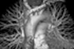
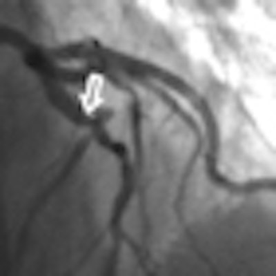
 Invasive coronary angiography and coronary CTA studies in a 62-year-old woman. Significant stenosis of the mid left anterior descending coronary artery (arrows) just proximal to the second diagonal branch was missed during initial interpretation of invasive coronary angiograms (upper left) due to superimposition of vessels. The lesion was detected on coronary CTA, displayed as curved multiplanar reformat (right). Re-evaluation of invasive coronary angiography (lower left) after unblinding of coronary CTA results confirms lesion in a single angiographic projection. Images courtesy of Dr. Matt Kerl and Dr. U. Joseph Schoepf.
On the other hand, 11 stenoses ≥ 50% in 11 patients seen at coronary CTA were not seen at the initial reading of angiography findings. After unblinding of CT findings, six of these stenoses were subsequently confirmed at angiography.
For example, in one patient conventional angiography missed left main artery stenosis, Schoepf said. Left main lesions "are notoriously difficult to evaluate with catheter angiography," he said, and in that patient "the tip of the catheter was seated beyond the lesion, so if you inject contrast in a spot distal to the actual lesion you will miss it." Catheter placement is, of course, not an issue in CT where you're getting a 3D view of the anatomy.
Per-patient results showed that coronary CTA had 98.2% (111/113) accuracy, 100% (45/45) sensitivity, and 97.1% (66/68) specificity, compared with 98.2% (111/113) accuracy, 95.6% (43/45) sensitivity, and 100% (68/68) specificity on conventional coronary angiography. The accuracies of the two exams did not differ significantly (p = 0.87).
Coronary CTA found significant coronary artery stenoses that had been missed initially on conventional angiography, and fortunately it missed only small lesions that were not amenable to intervention in any case, Kerl and colleagues wrote.
"Coronary CTA compares favorably with conventional coronary angiography for the diagnosis of patients with suspected coronary artery disease, although diagnostic accuracy still seems limited in smaller vessels and thus predominantly by spatial resolution," they wrote.
The composite reference standard used in this study "allowed controlling for false-negative conventional coronary angiography results by unblinding angiographers to coronary CTA findings and prompting re-evaluation of conventional coronary angiograms when results of the two tests were discrepant," the group wrote.
The typical radiation dose from coronary CTA was approximately 12 to 13 mSv, but in future studies the dose can be reduced substantially by moving to prospective gating for most patients, Schoepf said.
"I believe we have enough data and our own experience shows that you don't really lose anything in terms of diagnostic accuracy if you use prospective gating," Schoepf said. Even in patients who experience arrhythmia in the middle of an acquisition, most vendors have developed software that turns off the tube and pauses the acquisition until the extra systole has passed, such that arrhythmia is less and less an issue.
The study represents the first use of such an enhanced reference standard in cardiac imaging, but it's been successfully used in virtual colonoscopy, first by Dr. Perry Pickhardt from the University of Wisconsin-Madison in comparison with the conventional colonoscopy gold standard (New England Journal of Medicine, December 4, 2003, Vol. 349:23, pp. 2191-2220).
Pickhardt's use of an enhanced reference standard was definitely an inspiration for the study, Schoepf said, adding that an enhanced reference standard is the only way to analyze the performance of a new test on an level playing field.
"When we deal with novel technology that may be very good, we're always running up against the problem that we have to compare it to the gold standard," he said. But if even a flawed reference standard is the ideal, "an established methodology that's been around for years will always be superior," he said. But for a new test to have a chance, "it will have to exceed the performance of the reference standard," he said.
Angiography's limitations are well known, the authors said. "Because of its castlike nature, conventional coronary angiography detects coronary atherosclerotic disease only if the vascular lumen is compromised," they wrote.
Conventional coronary angiography shows cross-sectional anatomy from a 2D silhouette of the contrast-filled lumen. For a complicated coronary lesion, "arbitrary projection angles may significantly misrepresent stenosis severity," they wrote.
In addition, the subjective nature and strong observer dependence of conventional coronary angiography is well known.
Coronary CTA, on the other hand, "is a true cross-sectional imaging technique and thus not subject to the limitations of projection angiography," they wrote. The accumulation of atherosclerotic plaque is easily seen and can guide the reader to a better evaluation of affected vessel segments.
Intravascular ultrasound studies show that even significant plaque may not be depicted at angiography. However IVUS was not used in the study protocol and constitutes a study limitation, though IVUS is restricted to larger vessels.
Another limitation was the research setting, and the fact that all patients had been referred for angiography. The prevalence of disease was high at 39%, which tends to increase sensitivity.
Coronary CTA compares more favorably with conventional angiography than is suggested by the existing literature, the group concluded.
"For future studies, the use of an enhanced, composite reference standard would be more appropriate for assessing the true accuracy of coronary CTA for stenosis detection than comparison with conventional coronary angiography alone," they wrote. In clinical use, CT can even improve the performance of catheter angiography.
"For the next step, what we want to do is follow exactly the paradigm of virtual colonoscopy," which can be used to boost the results of conventional colonography in studies that perform both tests, he said.
So if coronary CTA shows significant findings -- for example, CT shows a left main stenosis at coronary CT that's difficult to visualize at angiography -- the radiologist can describe those results to the angiographer, who can acquire additional views "and either confirm or dismiss the finding" at angiography, Schoepf said.
"By integrating the CT results into catheter angiography you can improve the diagnostic yield and accuracy" of angiography, he said.