PET/CT imaging using gallium-68 (Ga-68) Fibroblast activation protein inhibitor (FAPI) radiotracer over F-18 FDG appears promising in women with invasive lobular carcinoma (ILC), according to a study published on 14 March in the Journal of Nuclear Medicine.
In an analysis of women diagnosed with ILC who underwent both imaging approaches, Ga-68 FAPI showed higher activity in tumors than F-18 FDG and identified more metastatic lesions, noted nuclear medicine physician and lead author Ertan Sahin, MD, of Gaziantep University in Gaziantep, Turkey.
“These insights can inform clinical decisions, providing a more comprehensive understanding of disease extent and refining treatment strategies for breast cancer patients,” the group wrote.
ILC is a subtype of breast cancer that begins in the milk-producing glands (lobules) of the breast and can spread to the lymph nodes and other areas. Women have a much poorer prognosis if the cancer spreads than if it is caught early. In diagnosed patients, accurate staging is vital for effective clinical management. And although F-18 FDG is a commonly used radiotracer in PET/CT imaging, its efficacy can vary among patients, according to the authors.
Conversely, FAPI (fibroblast activation protein inhibitor) radiotracers are believed to be more accurate and have shown promise in early studies in these patients. Yet more evidence is needed before it can be implemented clinically, the authors wrote.
“Although the benefits of Ga-68 FAPI-PET/CT have been demonstrated in numerous studies involving small patient cohorts, it is not yet routinely recommended by national guidelines because of the lack of high-level evidence,” the group wrote.
Thus, in this retrospective analysis, the researchers studied results in ILC-diagnosed women who underwent both F-18 FDG-PET/CT and Ga-68 FAPI-PET/CT imaging at their hospital in Gaziantep.
They included 23 women with a mean age of 51, with eight being premenopausal. Before undergoing PET/CT, four patients had undergone breast surgery. Additionally, two patients had bilateral breast cancer involvement.
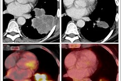
 Maximum-intensity projection F-18 FDG-PET (left) and Ga-68 FAPI-PET (right) images of synchronous ILC. (A) A 42-year-old patient with multicentric primary lesions (encircled), regional (solid arrows), and distant inferior jugular lymph node (dashed arrows) seen on F-18 FDG-PET and subsequently confirmed on Ga-68 FAPI-PET, which also revealed additional malignant foci aligned with MRI. Some lesions exhibited high activity retention on Ga-68 FAPI-PET. (B) A 53-year-old patient showing no significant uptake on F-18 FDG-PET but synchronous bilateral lesions (arrows) on Ga-68 FAPI-PET, which also identified pathologic lymph nodes (encircled), resulting in upstaging. Image courtesy of the Journal of Nuclear Medicine.
An analysis by two experienced nuclear medicine physicians revealed that Ga-68 FAPI-PET/CT outperformed F-18 FDG-PET/CT, with higher radiotracer uptake in primary tumors (p = 0.001). Moreover, Ga-68 FAPI-PET/CT scans revealed more metastatic cancer in axillary lymph nodes and identified more lesions, including bone and liver metastases, according to the results.
The authors noted that F-18 FDG-PET/CT remains a highly valuable tool. Both approaches identified an identical number of primary breast lesions. Nonetheless, more accurate staging of patients with ILC may improve how they are treated, as current imaging approaches have higher rates of false negatives, they wrote.
“This study underscores Ga-68 FAPI-PET/CT’s superiority over F-18 FDG-PET/CT for ILC,” the group concluded.
A link to the full study can be found here.