SINGAPORE - Understanding the relationship between proton density fat fraction and patient age in the thigh muscle could help clinicians identify specific ways to reduce functional decline associated with aging.
A team led by Jamie Scott, PhD, of Norwich Medical School, University of East Anglia in the U.K., found that increased fat fraction in the muscle is associated with decreased function as an individual ages. The group's research was presented at the International Society for Magnetic Resonance in Medicine (ISMRM) meeting.
Aging is associated with a progressive decline in skeletal muscle mass – also called sarcopenia – as well as a decline in muscle strength, the researchers explained, noting that "these two processes occur together, but the decline in strength outpaces the decline in mass by as much as a factor of five."
However, it's currently unclear which muscles may be affected first by this condition, and determining this could help clinicians better identify the onset of it, Scott and colleagues wrote. That's where MRI comes in: The modality is well-suited to the task of estimating intramuscular fat through chemical-shift-based water-fat-separation imaging, a method that has been shown to correlate with histology and is highly reproducible, making it "ideal for assessing muscle quality in longitudinal studies of aging," the authors noted.
Scott's group conducted a study that measured intramuscular fat in the thigh muscles of 94 healthy individuals (median age, 56), performing chemical-shift-based water-fat-separation MR imaging on a 3-tesla system to calculate proton density fat fraction (PDFF). The researchers assessed the quadriceps, the adductors, and the hamstrings and produced PDFF maps.
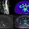
 Proton density fat fraction (PDFF) in the left thigh. Columns, from left to right, represent data acquired in 23-, 57-, and 89-year-old male participants, respectively. The top row shows T1-weighted images from the mid-thigh with posterior, medial, and anterior compartment segmentations overlaid. The middle row shows PDFF maps generated from Dixon data taken at the same position as the T1-weighted data; the color scale indicates PDFF values in percent. The bottom row shows density plots of pixelwise PDFF per compartment. Median PDFF increases with increasing age. Image and caption courtesy of coauthor Donnie Cameron, PhD, of Radboud University in Nijmegen, the Netherlands.
The group found the following:
- In the whole cohort, the median PDFF over the whole thigh was significantly associated with aging and increased fat fraction. The association between whole-thigh PDFF and age was stronger in women than in men.
- This finding held true for the quadriceps muscles but tended to be stronger in the hamstring muscles.
- The hamstring muscles biceps femoris short head and semimembranosus were most strongly associated with aging and increased fat fraction, while quadricep muscles sartorius and gracilis showed the weakest associations.
"We show that proton density fat fraction increases with age in the muscles of the thigh, particularly in women, and in the hamstring muscles -- suggesting these muscles may benefit most from interventions targeting sarcopenia," Scott and colleagues concluded.